
Page 11 - Human Environment Interface (3)
P. 11
Nanoparticle Behavior in Living Cells
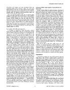
[YO] NPs in the medium, e.g. in the extracellular milieu, was Dynasore inhibits rapid uptake of nanoparticles into
significantly higher (DRICS.2 mm2s21; Figure 5H and J; area living cells
marked with m). These data are fully consistent with our FRAP
and FCS measurements and demonstrate the power of RICS to In order to further define the uptake mechanism a time-lapse of
spatially resolve NP dynamics and bio-interactions inside and living HEp-2 cells was recorded before and after the application of
outside of living cells with high precision. fluorescent PS NPs to the medium in the presence of dynasore.
Dynasore specifically inhibits the GTPase activity of dynamins,
Due to an improved signal to noise ratio in confocal images but does not interfere with other small GTPases [46]. Within 1–2
compared to single point FCS measurements, D values obtained seconds after addition it blocks all dynamin-dependent endocytic
for NPs in living cells by RICS can be determined with higher pathways by capturing clathrin-coated or uncoated pits in various
accuracy [23,42]. Notably, our data also reveal that RICS stages of vesicle formation before they pinch off the membrane
dynamics measurements of fluorescent particles within living cells [47]. Representative micrographs show that dynasore significantly
can be done in the presence of large immobile features commonly inhibits the cytoplasmic uptake of COOH-PS [YO] NPs
found in the cell (i.e. mitochondria). The accessible timescales in (Figure 7A). Quantification of intracellular NP-fluorescence shows
RICS span all the way from microseconds to seconds [23], as a 4-fold or 8-fold reduction, respectively, of signal intensity that is
evidenced by revealing a location-dependent 3 orders of magni- correlated to an increase of inhibitor concentration (Figure 7B).
tude difference in the diffusion coefficient of COOH-NPs in the Dynasore concentration-dependent decrease of cytoplasmic na-
cytoplasm (Figure 5J and K). nomaterial wash-in was likewise observed using differently labelled
COOH-PS [YG] NPs (Figure 7C). Since macropinocytosis is
Fast uptake of PS NPs into living cells dynamin-independent, the combination of our time kinetics and
To investigate the dynamics of cellular NP uptake, confocal inhibitor results suggests that PS NPs enter cells by fast, clathrin-
dependent or clathrin-independent endocytosis. The assumption
image series of living HEp-2 cells were recorded before, during of a clathrin-dependent portal for PS NP entry into cells is in
and after addition of fluorescent PS NPs to the medium (Figure 6). agreement with a recent report showing that in a monocytic cell
Fluorescence of COOH-PS [YO] NPs was detectable in the line anti-sense RNA-mediated depletion of dynamin II as well as
cytoplasm as early as 5 seconds after their addition to the cells clathrin results in significant inhibition of uptake of COOH- as
(Figure 6A). The subsequent time-lapse between 5 seconds and 1 well as amino-functionalized PS particles with a diameter of
minute is characterized by a steady increase of fluorescence 100 nm [48]. However, it has to be noted that the analyses by
intensity and accumulation of COOH-PS [YO] NPs in the Lunov et al. do not directly address fast uptake mechanisms, since
cytoplasm and at distinct reticulated structures resembling they were performed somewhat delayed, e.g. .30 minutes after
mitochondria. The particle-fluorescence in the cytoplasm and at addition of the particles.
mitochondria was quantitated in representative subcellular regions
of interest (Figure 6C). While accumulation of COOH-PS [YO] While PS as well as silica NPs rapidly traverse the cell
NPs in the cytoplasm reached a plateau after 30 seconds, membrane via dynamin-dependent endocytosis (Figure 7, and
accumulation at mitochondria continued to increase over several data not shown), COOH-PS [YO] NPs additionally showed rapid
minutes (Figure 6C). Ten minutes after particle addition no further endosomal escape by accumulation at mitochondria within a
increase in cellular fluorescence was observed (data not shown). minute. In contrast, plain-PS [YG] NPs recruit to and remain
The distinct fluorescence pattern and rapid uptake was likewise attached to vesicular structures in the perinuclear region. These
observed in HeLa cells and mouse embryonic fibroblasts (data not results are consistent with the idea that modulation of NP-
shown), indicating that cytoplasmic PS NP wash-in in the seconds properties can be developed into controlled subcellular targeting.
regimen is not cell-type specific. Applying the same imaging
settings, particle fluorescence began to appear in the nucleus 3 to 4 Conclusions
minutes after particle application (data not shown). This ’delay’ is
consistent with our FRAP data that revealed a relatively slow In this study we visualize the route of NP movement inside the
movement/exchange of nanoparticles at the nuclear membrane living cell in a time and space resolved manner. A protocol of
(Figure 3D). We next tested PS particles with different surface multimodal kinetic imaging is established that enables local and
characteristics, fluorochrome-labelling or size, and observed fast quantitative analyses of NP-behavior in live cells, e.g. a definition
uptake of plain-PS [YG] NPs into living cells (Figure 6D and E), of the subcellular NP-bio-interface. Given the existing expertise in
whereas bulk-sized COOH-PS [YG] particles with a diameter of fluorochrome-labelling of NPs or their intrinsic fluorescent
200 nm did not enter living HEp-2 cells during an observation properties, such systematic kinetic imaging has the potential to
time frame of 30 minutes (Figure 6F, and data not shown). develop a framework for definition of nanomaterial interactions at
the cell membrane and within subcellular compartments of living
The time kinetics of PS NP uptake correspond to pathways such cells. Our results show that location and interactions with cellular
as macropinocytosis and clathrin-dependent or clathrin-indepen- structures seem to correlate with distinct NP-characteristics, which
dent endocytosis where intracellular vesicles are observed after a implies that every (new) nanomaterial has to be tested individually.
few seconds to 1.5 minutes [9,43,44]. Caveolae can be excluded as However, it can be assumed that groups of pathways and schemes
portals of PS NP entry because in most cells caveolae are only of bio-interactions with common paradigms will emerge. Consis-
slowly internalized (halftime: 20 minutes) and the small vesicles tent with this concept, definition of individual cellular NP-
(50–60 nm in diameter) carry little fluid-phase volume [9]. In fact, networks may be instrumental in resolving as yet contradicting
the observation of COOH-PS [YO] NP occurring in the results concerning the fate of nanomaterials within living cells, and
cytoplasm within a few seconds after their addition to the cells respective cellular end points.
reminds of rapid rates of endocytosis that have been demonstrated
in nerve terminals with a time constant of t = 1–2 seconds [24,45]. Investigation of NP biophysics within living cells, as performed
While the underlying mechanisms are not yet fully understood, here, represents a prerequisite to better understand the molecular
e.g. if the endocytotic vesicles require a clathrin coat or form in a mechanisms of NP-bio-interactions, since this likewise requires
coat-independent manner, such events definitely are dynamin- quantitative analyses and observation of subcellular microenvi-
dependent [24]. ronments at high resolution. Elucidation of NP delivery within
PLOS ONE | www.plosone.org 9 April 2013 | Volume 8 | Issue 4 | e62018