
Page 142 - First Aid for the USMLE Step 1 2020, Thirtieth edition [MedicalBooksVN.com]_Neat
P. 142
98 SECTIon II Immunology ` Immunology—lymphoId StructureS Immunology ` Immunology—cellular componentS
Spleen Located in LUQ of abdomen, anterolateral to Splenic dysfunction (eg, postsplenectomy
left kidney, protected by 9th-11th ribs. state, sickle cell disease autosplenectomy):
A
Sinusoids are long, vascular channels in red IgM complement activation C3b
pulp (red arrows in A ) with fenestrated “barrel opsonization susceptibility to encapsulated
hoop” basement membrane. organisms.
T cells are found in the periarteriolar Postsplenectomy blood findings:
lymphatic sheath (PALS) within the white Howell-Jolly bodies (nuclear remnants)
pulp (white arrows in A ). Target cells
B cells are found in follicles within the Thrombocytosis (loss of sequestration and
white pulp. removal)
The marginal zone, in between the red pulp Lymphocytosis (loss of sequestration)
and white pulp, contains macrophages and Vaccinate patients undergoing splenectomy or
specialized B cells, and is where antigen- with splenic dysfunction against encapsulated
presenting cells (APCs) capture blood-borne organisms (pneumococci, Hib, meningococci).
antigens for recognition by lymphocytes.
Splenic macrophages remove encapsulated
bacteria.
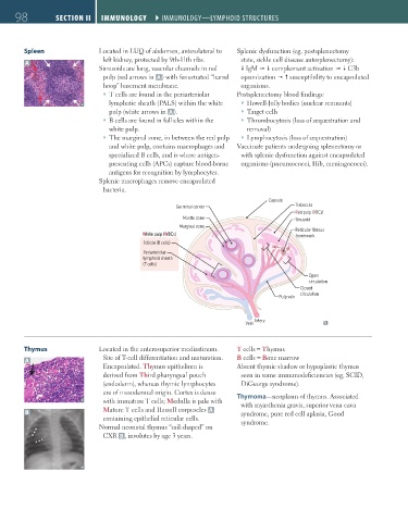
Capsule
Germinal center Trabecula
Red pulp (RBCs)
Mantle zone Sinusoid
Marginal zone
Reticular fibrous
White pulp (WBCs) framework
Follicle (B cells)
Periarteriolar
lymphoid sheath
(T cells)
Open
circulation
Closed
circulation
Pulp vein
Vein Artery
Thymus Located in the anterosuperior mediastinum. T cells = Thymus
A Site of T-cell differentiation and maturation. B cells = Bone marrow
Encapsulated. Thymus epithelium is Absent thymic shadow or hypoplastic thymus
derived from Third pharyngeal pouch seen in some immunodeficiencies (eg, SCID,
(endoderm), whereas thymic lymphocytes DiGeorge syndrome).
are of mesodermal origin. Cortex is dense Thymoma—neoplasm of thymus. Associated
with immature T cells; Medulla is pale with with myasthenia gravis, superior vena cava
B Mature T cells and Hassall corpuscles A syndrome, pure red cell aplasia, Good
containing epithelial reticular cells. syndrome.
Normal neonatal thymus “sail-shaped” on
CXR B , involutes by age 3 years.
FAS1_2019_02-Immunology.indd 98 11/7/19 3:24 PM