
Page 254 - First Aid for the USMLE Step 1 2020, Thirtieth edition [MedicalBooksVN.com]_Neat
P. 254
210 SECtIoN II Pathology ` PATHOLOGY—CeLLuLAr InjurY Pathology ` PATHOLOGY—CeLLuLAr InjurY
Ischemia Inadequate blood supply to meet demand. Mechanisms include arterial perfusion (eg,
A atherosclerosis), venous drainage (eg, testicular torsion, Budd-Chiari syndrome), shock.
Regions most vulnerable to hypoxia/ischemia and subsequent infarction:
OrGAn reGIOn
Brain ACA/MCA/PCA boundary areas a,b
Heart Subendocardium (LV) A
Kidney Straight segment of proximal tubule (medulla)
Thick ascending limb (medulla)
Liver Area around central vein (zone III)
a
Colon Splenic flexure (Griffith point), rectosigmoid
junction (Sudeck point) a
a Watershed areas (border zones) receive blood supply from most distal branches of 2 arteries with
limited collateral vascularity. These areas are susceptible to ischemia from hypoperfusion.
b Neurons most vulnerable to hypoxic-ischemic insults include Purkinje cells of the cerebellum and
pyramidal cells of the hippocampus and neocortex (zones 3, 5, 6).
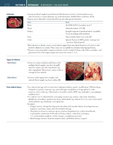
Types of infarcts
Red infarct Occurs in venous occlusion and tissues with A B
multiple blood supplies (eg, liver, lung A ,
intestine, testes), and with reperfusion (eg,
after angioplasty). Reperfusion injury is due to
damage by free radicals.
Pale infarct Occurs in solid organs with a single (end-
arterial) blood supply (eg, heart, kidney B ).
Free radical injury Free radicals damage cells via membrane lipid peroxidation, protein modification, DNA breakage.
Initiated via radiation exposure (eg, cancer therapy), metabolism of drugs (phase I), redox
reactions, nitric oxide (eg, inflammation), transition metals, WBC (eg, neutrophils, macrophages)
oxidative burst.
Free radicals can be eliminated by scavenging enzymes (eg, catalase, superoxide dismutase,
glutathione peroxidase), spontaneous decay, antioxidants (eg, vitamins A, C, E), and certain metal
carrier proteins (eg, transferrin, ceruloplasmin).
Examples:
Oxygen toxicity: retinopathy of prematurity (abnormal vascularization), bronchopulmonary
dysplasia, reperfusion injury after thrombolytic therapy
Drug/chemical toxicity: acetaminophen overdose (hepatotoxicity), carbon tetrachloride
(converted by cytochrome P-450 into CCl 3 free radical fatty liver [cell injury
apolipoprotein synthesis fatty change], centrilobular necrosis)
Metal storage diseases: hemochromatosis (iron) and Wilson disease (copper)
FAS1_2019_04-Pathol.indd 210 11/7/19 4:02 PM