
Page 648 - First Aid for the USMLE Step 1 2020, Thirtieth edition [MedicalBooksVN.com]_Neat
P. 648
604 SeCTIOn III Renal ` RENAL—PAthoLogy Renal ` RENAL—PAthoLogy
Renal cyst disorders
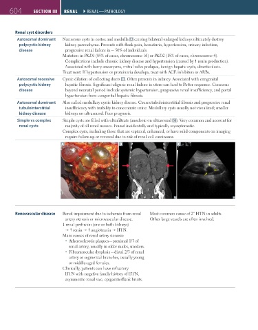
Autosomal dominant Numerous cysts in cortex and medulla A causing bilateral enlarged kidneys ultimately destroy
polycystic kidney kidney parenchyma. Presents with flank pain, hematuria, hypertension, urinary infection,
disease progressive renal failure in ~ 50% of individuals.
Mutation in PKD1 (85% of cases, chromosome 16) or PKD2 (15% of cases, chromosome 4).
Complications include chronic kidney disease and hypertension (caused by renin production).
Associated with berry aneurysms, mitral valve prolapse, benign hepatic cysts, diverticulosis.
Treatment: If hypertension or proteinuria develops, treat with ACE inhibitors or ARBs.
Autosomal recessive Cystic dilation of collecting ducts B . Often presents in infancy. Associated with congenital
polycystic kidney hepatic fibrosis. Significant oliguric renal failure in utero can lead to Potter sequence. Concerns
disease beyond neonatal period include systemic hypertension, progressive renal insufficiency, and portal
hypertension from congenital hepatic fibrosis.
Autosomal dominant Also called medullary cystic kidney disease. Causes tubulointerstitial fibrosis and progressive renal
tubulointerstitial insufficiency with inability to concentrate urine. Medullary cysts usually not visualized; smaller
kidney disease kidneys on ultrasound. Poor prognosis.
Simple vs complex Simple cysts are filled with ultrafiltrate (anechoic on ultrasound C ). Very common and account for
renal cysts majority of all renal masses. Found incidentally and typically asymptomatic.
Complex cysts, including those that are septated, enhanced, or have solid components on imaging
require follow-up or removal due to risk of renal cell carcinoma.
A B C
Renovascular disease Renal impairment due to ischemia from renal Most common cause of 2° HTN in adults.
artery stenosis or microvascular disease. Other large vessels are often involved.
renal perfusion (one or both kidneys)
renin angiotensin HTN.
Main causes of renal artery stenosis:
Atherosclerotic plaques—proximal 1/3 of
renal artery, usually in older males, smokers.
Fibromuscular dysplasia—distal 2/3 of renal
artery or segmental branches, usually young
or middle-aged females.
Clinically, patients can have refractory
HTN with negative family history of HTN,
asymmetric renal size, epigastric/flank bruits.
FAS1_2019_14-Renal.indd 604 11/7/19 5:42 PM