
Page 698 - First Aid for the USMLE Step 1 2020, Thirtieth edition [MedicalBooksVN.com]_Neat
P. 698
654 SectioN iii RepRoductive ` REPRODUCTIVE—PATHOlOgy RepRoductive ` REPRODUCTIVE—PHARmACOlOgy
Epididymitis and Most common causes:
orchitis C trachomatis and N gonorrhoeae (young men)
E coli and Pseudomonas (elderly, associated with UTI and BPH)
Autoimmune (eg, granulomas involving seminiferous tubules)
Epididymitis Inflammation of epididymis. Presents with localized pain and tenderness over posterior testis.
⊕ Prehn sign (pain relief with scrotal elevation). May progress to involve testis.
Orchitis Inflammation of testis. Presents with testicular pain and swelling. Mumps orchitis infertility risk.
Rare in boys < 10 years old.
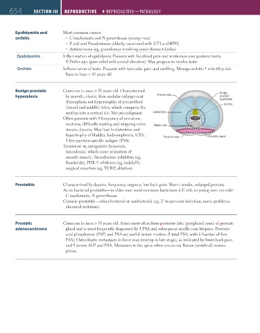
Benign prostatic Common in men > 50 years old. Characterized Benign
hyperplasia by smooth, elastic, firm nodular enlargement Anterior lobe prostatic
(hyperplasia not hypertrophy) of periurethral hyperplasia
(lateral and middle) lobes, which compress the Urethra
urethra into a vertical slit. Not premalignant. Lateral lobe
Often presents with frequency of urination,
nocturia, difficulty starting and stopping urine Middle lobe
stream, dysuria. May lead to distention and
hypertrophy of bladder, hydronephrosis, UTIs. Posterior lobe Prostate cancer
free prostate-specific antigen (PSA).
Treatment: α -antagonists (terazosin,
1
tamsulosin), which cause relaxation of
smooth muscle; 5α-reductase inhibitors (eg,
finasteride); PDE-5 inhibitors (eg, tadalafil);
surgical resection (eg, TURP, ablation).
Prostatitis Characterized by dysuria, frequency, urgency, low back pain. Warm, tender, enlarged prostate.
Acute bacterial prostatitis—in older men most common bacterium is E coli; in young men consider
C trachomatis, N gonorrhoeae.
Chronic prostatitis—either bacterial or nonbacterial (eg, 2° to previous infection, nerve problems,
chemical irritation).
Prostatic Common in men > 50 years old. Arises most often from posterior lobe (peripheral zone) of prostate
adenocarcinoma gland and is most frequently diagnosed by PSA and subsequent needle core biopsies. Prostatic
acid phosphatase (PAP) and PSA are useful tumor markers ( total PSA, with fraction of free
PSA). Osteoblastic metastases in bone may develop in late stages, as indicated by lower back pain
and serum ALP and PSA. Metastasis to the spine often occurs via Batson (vertebral) venous
plexus.
FAS1_2019_15-Repro.indd 654 11/7/19 5:52 PM