Page 295 - Hall et al (2015) Principles of Critical Care-McGraw-Hill
P. 295

CHAPTER 28: Interpretation of Hemodynamic Waveforms  199


                             A.                                     B.


                                Pra                                   Pra
                                                                    60
                             30                                                         v     v      v
                                       v    v     v
                                    a
                                     x
                                         y
                                                                    0
                              0

                                                    C.


                                                     Pra


                                                             v
                                                 30 –      c


                                                              Y
                                                           INSP                 INSP
                                                  0 –
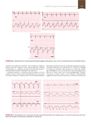
                    FIGURE 28-26.  Right atrial pressure (Pra) tracings in patients with tricuspid regurgitation. A and B. prominent v waves. C. broad c-v wave with large y descent. Scale in millimeters of mercury.


                    consistent with pulmonary embolism. One hemodynamic difference   differentiate atrial flutter from sinus tachycardia and paroxysmal supra-
                    between  these  two  conditions  is  that  massive  pulmonary  embolism   ventricular tachycardia, even with the aid of a 12-lead ECG. Although
                                                                      33
                    is characterized by a significant increase in the Ppad-Ppw gradient,    the response to adenosine is often the best way to define the underly-
                    whereas the latter should be unaffected by RV infarction. 50  ing atrial rhythm, in some cases atrial flutter may also be diagnosed by
                                                                                                                     29
                     Arrhythmia evaluation is sometimes aided by analysis of the Pra   detection of “flutter” waves in the Pra tracing (Fig. 28-28).  Similarly,
                    waveform. Narrow-complex, regular tachyarrhythmias at a rate of 140 to    the presence of regular cannon  a waves during the tachyarrhythmia
                    180 beats per minute are common in the ICU. It is sometimes difficult to   suggests atrioventricular dissociation due to a reentrant supraventricular


                                                           ECG
                                                                                                                ECG


                                                             Part
                             100                                                                                Part
                                                                        150


                                                             Pra
                              30                                        30                                      Pra
                                    y      y     y
                                   x     x      x
                                                                              x   y    x   y   x   y


                    FIGURE 28-27.  Right atrial pressure (Pra) tracing in patient with cardiac tamponade showing blunted y descent with preservation of x descent. After pericardiocentesis, the y descent
                    becomes more prominent. See text for discussion of physiology. Part, arterial pressure.


            section02.indd   199                                                                                       1/13/2015   2:05:42 PM
   290   291   292   293   294   295   296   297   298   299   300