
Page 290 - Hall et al (2015) Principles of Critical Care-McGraw-Hill
P. 290
194 PART 2: General Management of the Patient
in systolic Ppa by the change in alveolar pressure (plateau pressure—
Pra or Ppw PEEP) during a controlled tidal breath, with the change in Ppa reflecting
40
20 the change in Ppl. Next, PEEP is multiplied by the transmission ratio to
estimate end-expiratory Ppl. Finally, transmural pressure is calculated by
subtracting Ppl from the Pra or Ppw. Even though this method appears
40
10
40
Pressure Ppl to yield a valid estimate of transmural pressure, it is unclear whether
it contributes significantly to patient management. In clinical decision
making, use of the Ppw or Pra should not focus excessively on its abso-
0
lute value. It is often more important to assess how a change in the Ppw
or Pra correlates with clinically relevant clinical end points (eg, blood
−10 pressure, cardiac output, oxygenation, urine output) after manipulation
of intravascular volume, and this can be assessed without correcting for
the effect of PEEP.
The effect of PEEP on transmural pressure described above is relevant
FIGURE 28-16. Effect of changes in pleural pressure (Ppl) on the right atrial (Pra) or to both the Pra and Ppw. There is a second way in which PEEP may influ-
wedge pressure (Ppw) during assisted mechanical ventilation. Negative deflections in Ppl and
Pra/Ppw result from inspiratory muscle activity, and subsequent positive deflections represent ence the Ppw—but not the Pra. This mechanism involves compression
of the pulmonary microvasculature at high levels of PEEP that inter-
lung inflation by the ventilator. Pressure at end expiration (arrow) gives the best estimate of
transmural pressure. Scale in millimeters of mercury. rupts the continuous column of blood between the catheter tip and left
atrium, resulting in a Ppw that reflects alveolar rather than pulmonary
venous pressure. Fortunately, this phenomenon appears to be rare. High
ranging from 24% to 37% in one study. Conversely, decreased chest levels of applied PEEP are generally restricted to patients with severe
39
wall compliance due to intra-abdominal hypertension or morbid obesity ARDS and damaged lungs do not transmit alveolar pressure as fully to
41
will increase the percentage of PEEP transmission, as may be suggested the capillary bed as do normal lungs. A study of patients with ARDS
by large swings in intrathoracic vascular pressure during tidal ventila- demonstrated that the Ppw faithfully reflected simultaneously measured
42
tion (Fig. 28-18). Auto-PEEP may have a greater impact on transmural LVEDP even at a PEEP of 16 to 20 cm H O. Concern that the Ppw may
2
pressures than an equivalent degree of applied PEEP, because auto-PEEP represent alveolar pressure should be restricted to those rare instances
usually occurs in the setting of normal or increased lung compliance, in which the Ppw tracing has an unnaturally smooth appearance that is
allowing a larger component of the alveolar pressure to be transmitted uncharacteristic of an atrial waveform, the Ppw approximates 75% of
to the juxtacardiac space. the applied PEEP (1 cm H O ~ 0.74 mm Hg), and the change in Ppw is
2
The effect of PEEP on transmural pressures can be reliably estimated significantly greater than the change in systolic Ppa (reflecting change in
in patients with a PAC who are undergoing controlled mechanical ven- Ppl) during a controlled ventilator breath. 43
pleural space (the transmission ratio) is calculated by dividing the change ■ ACTIVE (FORCED) EXPIRATION
tilation. First, the fraction of alveolar pressure that is transmitted to the
Contraction of abdominal expiratory muscles increases intrathoracic pres-
sure at end expiration. In contrast to PEEP, the increased intra-abdominal
pressure generated by expiratory muscles is almost fully transmitted to the
Alveolus pleural space. 44,45 Forceful expiration typically leads to a far greater overesti-
0 Left atrium
mation of transmural pressure than does the application of PEEP. Previous
studies have shown that forced expiration often causes the end-expiratory
16 Ppw to overestimate transmural pressure by more than 10 mm Hg. 24-26,45,46
−2 Given this magnitude of error, failure to appreciate forced exhalation as
Pleural space
15 the cause of an elevated Ppw or Pra may lead to inappropriate treatment of
hypovolemic patients with diuretics or vasopressors.
16
In mechanically ventilated patients, sedation (or even paralysis) may
+4 be used to reduce or eliminate expiratory muscle activity (Fig. 28-19). 25,26
In the nonintubated patient, recording the pressure tracing while the
PEEP Pleural pressure Intracardiac pressure Transmural pressure patient sips water through a straw sometimes helps eliminate large respi-
cm H O mm Hg mm Hg mm Hg ratory fluctuations (Fig. 28-19). An esophageal balloon has been used
45
2
to better estimate transmural pressure, but placement of esophageal
24
0 −2 16 18 catheters may not be well received by the dyspneic patient. A simpler
15 +4 16 12
method is to subtract the expiratory rise in bladder pressure from the
FIGURE 28-17. The effect of positive end-expiratory pressure (PEEP) on transmural pressure. end-expiratory Pra to obtain a “corrected” value to estimate transmural
45
In this example, 50% of PEEP is transmitted to the juxtacardiac space (15 cm H O ~ 12 mm Hg). pressure (Fig. 28-20). In two studies that used this approach, there was
2
Pra
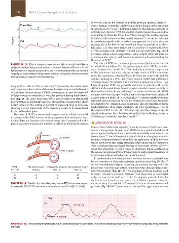
30
24 mm Hg
4 mm Hg
0
FIGURE 28-18. The large change in right atrial pressure (Pra) during mechanical ventilation reflects a marked increase in pleural pressure due to very low chest wall compliance. mm Hg, millimeters
of mercury.
section02.indd 194 1/13/2015 2:05:36 PM