
Page 292 - Hall et al (2015) Principles of Critical Care-McGraw-Hill
P. 292
196 PART 2: General Management of the Patient
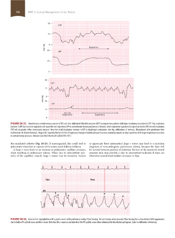
30
CVP
15
mm Hg 0 Expiration
30
IAP
15
0
30
CVP
15
mm Hg 30 0
IAP
15
Inspiration Expiration
0
FIGURE 28-21. Simultaneous central venous pressure (CVP) and intra-abdominal (bladder) pressure (IAP) tracings in two patients with large respiratory excursions in CVP. Top, expiratory
increase in IAP due to active expiration will cause the end-expiratory CVP to overestimate transmural pressure. Bottom, when expiration is passive (no expiratory rise in IAP) the end-expiratory
CVP will accurately reflect transmural pressure. Note the small inspiratory increase in IAP to diaphragm contraction. mm Hg, millimeters of mercury. (Reproduced with permission from
Leatherman JW, Bastin-DeJong C, Shapiro RS, Saavedra-Romero R. Use of expiratory change in bladder pressure to assess expiratory muscle activity in patients with larger respiratory excursions
in central venous pressure. Intensive Care Med. March 2012;38(3):453-457.)
the uninflated catheter (Fig. 28-23). If unrecognized, this could lead to to appreciate these intermittent large v waves may lead to a mistaken
pulmonary infarction or rupture of the artery upon balloon inflation. diagnosis of noncardiogenic pulmonary edema, because the Ppw will
A large v wave leads to an increase in pulmonary capillary pressure, be normal between periods of ischemia. Review of the monitor’s stored
often resulting in pulmonary edema. When due to intermittent isch- pressure data may provide a clue to intermittent ischemia if there are
emia of the papillary muscle, large v waves may be transient. Failure otherwise unexplained sudden increases in Ppa.
Ppa Ppw
60
S S V
V V V V
FIGURE 28-22. Acute mitral regurgitation with a giant v wave in the pulmonary wedge (Ppw) tracing. The pulmonary artery pressure (Ppa) tracing has a characteristic bifid appearance
due to both a PA systolic wave and the v wave. Note that the v wave occurs later than the PA systolic wave when referenced to the electrocardiogram. Scale in millimeters of mercury.
section02.indd 196 1/13/2015 2:05:38 PM