
Page 291 - Hall et al (2015) Principles of Critical Care-McGraw-Hill
P. 291
CHAPTER 28: Interpretation of Hemodynamic Waveforms 195
A B Baseline
Baseline 20 Pra 18
– 37.5 –
– 25 –
Ppw ~ 28 – 12.5 –
– 0 –
0
Pra 10 Sipping
20
– 25 – Postparalysis
– 12.5 –
– 0 – Ppw ~ 28
0
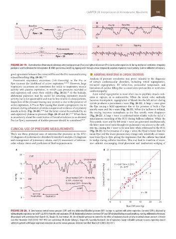
FIGURE 28-19. Overestimation of transmural pulmonary artery wedge pressure (Ppw) and right atrial pressure (Pra) due to active expiration. A. During mechanical ventilation, temporary
paralysis is used to eliminate forced expiration. B. With spontaneous breathing, sipping water through a straw temporarily suspends respiratory muscle activity. Scale in millimeters of mercury.
good agreement between the corrected Pra and the Pra measured during ■ ABNORMAL WAVEFORMS IN CARDIAC DISORDERS
relaxed breathing (Fig. 28-20). 45,46 Analysis of pressure waveforms may prove valuable in the diagnosis
Prominent respiratory excursions (>8-10 mm Hg) in the Pra or of certain cardiovascular disorders, including mitral regurgitation,
Ppw increase the likelihood of active expiration. 25,26,46 However, large tricuspid regurgitation, RV infarction, pericardial tamponade, and
respiratory excursions are sometimes due solely to inspiratory muscle limitation of cardiac filling due to constrictive pericarditis or restrictive
activity with passive expiration, in which case pressures recorded at cardiomyopathy.
end expiration will retain their validity (Fig. 28-21). At the bedside, Acute mitral regurgitation is most often due to papillary muscle isch-
46
abdominal palpation may be useful for detecting expiratory muscle emia or rupture, or to endocarditis. When the mitral valve suddenly
activity, but is not quantitative and may be less reliable in obese patients. becomes incompetent, regurgitation of blood into the left atrium during
Inspection of the pressure tracing may provide a clue to the presence of systole produces a prominent v wave (Fig. 28-22). A large v wave gives
active expiration. A Pra or Ppw tracing that shows a progressive rise in the Ppa tracing a bifid appearance due to the presence of both a Ppa
pressure during exhalation provides unequivocal evidence of expiratory systolic wave and the v wave (Fig. 28-22). When the balloon is inflated,
muscle activity (Fig. 28-20), 27,46 but the latter cannot be excluded by an the tracing becomes monophasic as the Ppa systolic wave disappears
end-expiratory plateau in pressure (Figs. 28-19 and 21). 46,47 When there (Fig. 28-22). A large v wave is confirmed most reliably with the aid of a
is uncertainty about the contribution of forced exhalation to an elevated simultaneous recording of the ECG during balloon inflation. While the
Pra (or Ppw), assessment of bladder pressure should be considered. 45,46 Ppa systolic wave and the left atrial v wave are generated simultaneously,
the latter must travel back through the pulmonary vasculature to the cath-
CLINICAL USE OF PRESSURE MEASUREMENTS eter tip, causing the v wave to be seen later when referenced to the ECG
(Fig. 28-22). In the presence of a large v wave, the Ppad is lower than the
There are three principal uses of intravascular pressures in the ICU: mean Ppw and the mean pressure may change only minimally on transi-
(1) diagnosis of cardiovascular disorders by waveform analysis, (2) diagnosis tion from Ppa to Ppw, giving the impression that the catheter has failed
and management of pulmonary edema, and (3) assessment of intravas- to wedge during catheter insertion. This may lead to insertion of exces-
cular volume status and prediction of fluid responsiveness. sive catheter, encouraging distal placement and inadvertent wedging of
A B 25
30
CVP
Uncorrected 20
CVP
15 Y = 0.6719x + 3.3313
R = 0.77
0 Corrected CVP (mm Hg) 15
mm Hg 30 IAP Uncorrected CVP = 18 mm Hg 10
IAP = 9 mm Hg
Corrected CVP = 9 mm Hg
15 IAP
5
n = 36
Inspiration Expiration
0
0
0 5 10 15 20 25
Best CVP (mm Hg)
FIGURE 28-20. A. Simultaneous central venous pressure (CVP) and intra-abdominal (bladder) pressure (IAP) tracings in a patient with active expiration. Corrected CVP is obtained by
subtracting the expiratory rise in IAP (Δ IAP) is from the end-expiratory CVP. B. Relationship between corrected CVP and CVP obtained during relaxed breathing. mm Hg, millimeters of mercury.
(Reproduced with permission from Qureshi AS, Shapiro RS, Leatherman JW. Use of bladder pressure to correct for the effect of expiratory muscle activity on central venous pressure. Intensive
Care Med. November 2007;33(11):1907-1912 and Leatherman JW, Bastin-DeJong C, Shapiro RS, Saavedra-Romero R. Use of expiratory change in bladder pressure to assess expiratory muscle
activity in patients with larger respiratory excursions in central venous pressure. Intensive Care Med. March 2012;38(3):453-457.)
section02.indd 195 1/13/2015 2:05:37 PM