
Page 294 - Hall et al (2015) Principles of Critical Care-McGraw-Hill
P. 294
198 PART 2: General Management of the Patient
II 0.5-40 Hz
1 mV
30 Ppw
24
18
12
6
0
AO
LA
LV
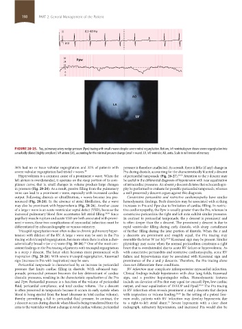
FIGURE 28-25. Top, pulmonary artery wedge pressure (Ppw) tracing with small v waves despite severe mitral regurgitation. Bottom, left ventriculogram shows severe regurgitation into
a markedly dilated (highly compliant) left atrium (LA), accounting for the minimal pressure change (small v wave). LV, left ventricle; AO, aorta. Scale in millimeters of mercury.
36% had no or trace valvular regurgitation and 32% of patients with pressure is therefore unaffected. As a result, there is little (if any) change in
severe valvular regurgitation had trivial v waves. 49 Pra during diastole, accounting for the characteristically blunted y descent
Hypervolemia is a common cause of a prominent v wave. When the of pericardial tamponade (Fig. 28-27). 29,50 Attention to the y descent may
left atrium is overdistended, it operates on the steep portion of its com- be useful in the differential diagnosis of hypotension with near equalization
pliance curve; that is. small changes in volume produce large changes of intracardiac pressures. An absent y descent dictates that echocardiogra-
in pressure (Fig. 28-24). As a result, passive filling from the pulmonary phy be performed to evaluate for possible pericardial tamponade, whereas
veins can lead to a prominent v wave, especially with increased cardiac a well-preserved y descent argues against this diagnosis.
output. Following diuresis or ultrafiltration, v waves become less pro- Constrictive pericarditis and restrictive cardiomyopathy have similar
nounced (Fig. 28-24). In the absence of atrial fibrillation, the a wave hemodynamic findings. Both disorders may be associated with striking
may also be prominent with hypervolemia (Fig. 28-24). Another cause increases in Pra and Ppw due to limitation of cardiac filling. In restric-
of a large v wave is an acute ventricular septal defect (VSD), because the tive cardiomyopathy, the Ppw is usually greater than the Pra, whereas in
increased pulmonary blood flow accentuates left atrial filling. 29,50 Since constrictive pericarditis the right and left atria exhibit similar pressures.
papillary muscle rupture and acute VSD are both associated with promi- In contrast to pericardial tamponade, the y descent is prominent and
nent v waves, these two complications of myocardial infarction must be is often deeper than the x descent. The prominent y descent is due to
differentiated by echocardiography or venous oximetry. rapid ventricular filling during early diastole, with sharp curtailment
Tricuspid regurgitation most often is due to chronic pulmonary hyper- of further filling during the later portion of diastole. When the x and
tension with dilation of the RV. A large v wave may be seen in the Pra y descents are prominent and roughly equal, the Pra tracing may
tracing with tricuspid regurgitation, but more often there is often a char- resemble the letter W (or M). 29,50 Kussmaul sign may be present. Similar
acteristically broad v (or c-v) wave (Fig. 28-26). One of the most con- physiology may occur when the normal pericardium constrains a right
29
sistent findings in the Pra tracing of patients with tricuspid regurgitation heart that is overdistended due to acute RV failure or hypervolemia. As
is a steep y descent. The latter often becomes more pronounced with with constrictive pericarditis and restrictive cardiomyopathy, acute RV
inspiration (Fig. 28-26). With severe tricuspid regurgitation, Kussmaul failure and hypervolemia may be associated with Kussmaul sign and
sign (increase in Pra with inspiration) may be seen. prominence of the x and y descents. Therefore, the Pra tracing alone
Pericardial tamponade is characterized by an increase in pericardial does not differentiate these conditions.
pressure that limits cardiac filling in diastole. With advanced tam- RV infarction may complicate inferoposterior myocardial infarction.
ponade, pericardial pressure becomes the key determinant of cardiac Clinical findings include hypotension with clear lung fields, Kussmaul
diastolic pressures, resulting in the characteristic equalization of the Pra sign, and a positive hepatojugular reflux. Hemodynamic features
and Ppw. Pericardial pressure is a function of the volume of pericardial include an elevation of Pra that may equal (or exceed) Ppw, low cardiac
fluid, pericardial compliance, and total cardiac volume. The x descent output, and near equalization of RVEDP and Ppad. 29,50 The Pra tracing
is often preserved in tamponade because it occurs in early systole when in RV infarction often reveals prominent x and y descents that deepen
blood is being ejected from the heart (decrease in total cardiac volume), with inspiration or volume loading. 29,50 In the setting of a patent fora-
thereby permitting a fall in pericardial fluid pressure. In contrast, the men ovale, patients with RV infarction may develop hypoxemia due
y descent occurs during diastole when blood is being transferred from the to a right-to-left atrial shunt. Severe hypoxemia with a clear chest
51
atria to the ventricles without a change in total cardiac volume; pericardial radiograph, refractory hypotension, and increased Pra would also be
section02.indd 198 1/13/2015 2:05:41 PM