
Page 374 - Hall et al (2015) Principles of Critical Care-McGraw-Hill
P. 374
244 PART 3: Cardiovascular Disorders
), and effective O extraction by the tissues before
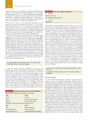
2 TABLE 32-2 Reasons For Sv O 2 and Scv O 2 To Be Dissimilar
oxygen consumption (V O 2
using Scv O 2 or Sv O 2 as markers of circulatory sufficiency. D O 2 is the product
is the sum of oxygen bound to hemoglo- Scv O 2 > Sv O 2
of cardiac output and Ca O 2 . Ca O 2
) and dissolved Normal (usually ~2%-3%)
bin (product of hemoglobin concentration [Hb] and Sa O 2
+
oxygen (Pa O 2 ). Although the formula defining Ca O 2 is Hb·1.36·Sa O 2 Intra-abdominal compartment syndrome
·0.0031, the amount of O dissolved in the plasma is minimal except
Pa O 2 2
under extreme hyperbaric conditions, and is usually ignored. Sv O 2 > Scv O 2
Circulatory shock
must equal cardiac output times the dif-
Clinical Uses of Sv O 2 : Since V O 2
remains
2
ference in Ca O 2 and mixed venous O content (Cv O 2 ), if Ca O 2
will vary in proportion to cardiac output. measuring a mixed venous sample of blood at that site, Scv O 2 reflects
relatively constant then Cv O 2
can be easily measured and Hb rarely upper body venous blood while ignoring venous drainage from the
Recall from above that Sp O 2
changes rapidly, thus in the sedated patient without changing stress, this lower body (eg, intra-abdominal organs). Accordingly, Scv O 2 is usu-
assumption is true. Since the amount of O dissolved in the plasma is ally higher than Sv O 2 by 2% to 3% in a sedated resting patient because
2 cerebral O consumption is minimal and always sustained above other
very small, the primary factor determining changes in Cv O 2 will be Sv O 2 . 2
correlates well with the O supply-to-demand ratio. 14 because the
Thus, Sv O 2 2 organs. Still, with agitation Scv O 2 can become less than Sv O 2
However, several relevant conditions may limit this simple appli- lower body extracts less O than the upper body, making inferior vena
2
16
in assessing circulatory sufficiency and cardiac output caval O saturation higher. Different vascular beds have different
cation of Sv O 2 2
were to increase (as occurs with exercise), hemoglobin- venous O saturations owing to their different functions. The kidneys
(Table 32-1). If V O 2 2
carrying capacity to decrease (as occurs with anemia, hemoglobin- have an extremely high venous O saturation because they function
2
to decrease (as occurs with more to ultrafilter plasma than to extract O . In circulatory shock,
opathies, and severe hemorrhage), or Sp O 2 2
renal blood flow markedly decreases, such that inferior vena caval O
hypoxic respiratory failure), then for the same cardiac output, Sv O 2 2
would also decrease. Similarly, if more blood flows through nonmeta- saturation decreases markedly. During cardiogenic or hypovolemic
bolically extracting tissues as occurs with intravascular shunts, or mito- shock, mesenteric and renal blood flow decreases, thus increasing
17
chondrial dysfunction limits O uptake by tissues (as may occur with local O extraction. In septic shock, splanchnic O consumption
2
2
2
carbon monoxide poisoning and potentially in prolonged sepsis), then increases, thus increasing local O extraction despite increased cardiac
2
18
even though output. Finally, since muscle is highly efficient at extracting O ,
Sv O 2 will increase for a constant cardiac output and V O 2 2
circulatory stress exists and may cause organ dysfunction. Intravascular muscular activity results in a marked decrease in its venous O satu-
2
shunts and mitochondrial dysfunction are the purported causes of high ration. Depending on whether the activity is in the upper extremities
in patients with fluid-resuscitated septic shock. Whether there is (superior vena cava), lower extremities (inferior vena cava), or trunk
Sv O 2
mitochondrial dysfunction in sepsis is unclear but there is good evi- (azygous), Sv O 2 and Scv O 2 can change differently. Thus, Scv O 2 cannot
dence of microvascular shunting. 15 be used as surrogate for Sv O 2 under conditions of circulatory shock. If
probably exists, but
19
Scv O 2 is <65%, however, then inadequate D O 2
is >70% it has no prognostic utility. 20,21 Still, when the whole
if Scv O 2
defines increased change
Sv O 2 is the gold standard for assessing circulatory stress. A low Sv O 2 body O supply/O demand ratio is altered, both Sv O 2 and Scv O 2
2
2
circulatory stress, which may or may not be pathological. in similar directions. 22
has covary in the extremes but may change in opposite directions as condi-
Sv O 2 and Scv O 2 : Recent interest in estimating Sv O 2 using Scv O 2 Sv O 2 and Scv O 2
increased with the lack of enthusiasm for insertion of pulmonary tions change.
arterial catheters. Since central venous catheterization is commonly threshold values to define circulatory stress are only relevant if low (a high Scv is
performed as a stable access site for fluid and drug infusion, direct Scv O 2 O 2
nondiagnostic).
access to central venous blood is also commonly available in most
does not sample true mixed
critically ill patients. However, Scv O 2 ■
venous blood and most vena caval blood flow is laminar, thus if the TISSUE OXIMETRY
tip is in one of these laminar flow sites it will preferentially report The most currently used technique to measure peripheral tissue O
a highly localized venous drainage site O saturation. Clearly, the 2
2 saturation (St O 2 ) is near-infrared spectroscopy (NIRS) (InSpectre,
potential exists for spurious estimates of Sv O 2 (Table 32-2). Most Hutchinson Industries, Hutchinson, MN). NIRS is a noninvasive tech-
central venous catheters are inserted from internal jugular or sub- nique based in the differential absorption properties of oxygenated and
clavian venous sites with their distal tip residing in the superior deoxygenated hemoglobin to assess the muscle oxygenation. Using non-
vena cava, usually about 5 cm above the right atrium. Thus, even if infrared light (680-800 nm) that is mostly absorbed in the tissue by the
hemoglobin, the signal receptor is able to quantify the amount of oxy-
genated hemoglobin present in the tissue crossed by the near-infrared
to Trend Circulatory Sufficiency
light. Only the small vessels like arterioles or capillaries are monitored
TABLE 32-1 Limitations to the Use of Sv O 2
independent of cardiac output with this technique, as the big vessels like veins or arteries have too
Independent events that decrease Sv O 2
Event Process much concentration with blood and absorb the light preventing photon
emergence. Different commercially available devices have different tissue
Exercise
penetration levels and sensitivities to changes in hemoglobin concentra-
Increase V O 2
Anemia Decreased O -carrying capacity tions. A major focus of NIRS device evaluation is in the assessment of
2
Hypoxemia Decreased arterial O content cerebral ischemia, which is beyond the scope of this chapter. Still, all
2
independent of cardiac output NIRS devices tend to display similar performance in assessing local St O 2 .
Independent events that increase Sv O 2 measures have been studied to assess tissue
Noninvasive NIRS St O 2
Event Process hypoperfusion in different populations. 23,24 Although there is a good
Sepsis Microvascular shunting correlation between the absolute St O 2 value and some other cardiovas-
End-stage hepatic failure Macrovascular shunting cular indexes, 25,26 the capacity of the baseline St O 2 values to identify
impending cardiovascular insufficiency is limited (sensitivity, 78%;
Carbon monoxide poisoning Mitochondrial respiratory chain inhibition
specificity, 39%) and no more accurate than a single systolic blood
section03.indd 244 1/23/2015 2:06:49 PM