
Page 370 - Hall et al (2015) Principles of Critical Care-McGraw-Hill
P. 370
240 PART 3: Cardiovascular Disorders
lower than Cp in cardiogenic edema. When the vascular membrane is
77
Alveolus repaired, alveolar edema is cleared very slowly from noninjured lungs by
active transport of sodium; water follows the osmotic gradient through an
intact alveolar membrane, and this clearance raises alveolar protein con-
Pmv mv
centration above Cp as a clinical marker of recovery from ARDS. 86
Pis is LVEDP PEEP increases end-expired lung volume to decrease Pis and increase
capacity in the peribronchovascular interstitium; this in turn redistrib-
Alveolus
utes much of the alveolar edema into this interstitial reservoir, associated
with the aeration of flooded airspaces at a much larger alveolar volume
Edema flow = [ (Pmv - Pis) - ( mv - is) ]K f to reduce shunt and to increase lung compliance without altering the
amount of edema. 13,14,87 Because lung volume increases greatly when
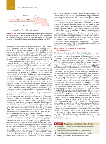
FIGURE 31-12. Schematic representation of Starling forces governing the flux of lung liquid PEEP is effective in redistributing edema, Ppl must increase to push the
from the intravascular to the extravascular space (for discussion, see text). is, interstitial space; chest wall to an equivalently higher volume. This raises Pra to reduce VR
LVEDP, left ventricular end-diastolic pressure; mv, microvessels of the lung; π, colloid osmotic and BP 5,6,26 unless the patient’s baroreceptor reflexes, iatrogenic infusions
30
pressure; σ, reflection coefficient. (Reproduced with permission from Hall and Wood LDH ) of fluid, or vasoactive drugs maintain Pms and Q ˙ t. 31,88 This recruitment
of previously flooded airspaces occurs within the large P-V hysteresis of
the edematous lung, so less PEEP is required than that indicated by the
inflection point of the inflation P-V curve. 89
albumin and globulin. If these plasma proteins were completely reflected ■
(σ = 1), no protein would pass from lung blood to the interstitium; in AN APPROACH TO MANAGING ACUTE HYPOXEMIC
contrast, if the microvascular membrane were freely permeable (σ = 0), RESPIRATORY FAILURE
interstitial protein concentration (Cl), as measured in lung lymph, would As with many therapeutic interventions in critical illness, too much
equal that of plasma proteins (Cp). C /C is about 0.6 in the normal can cause harm, so it is helpful to define the goal of each intervention
p
l
steady-state edema flow in most mammals; when Q ˙ e, as estimated from and then use the mildest intervention to achieve that goal. Ventilator
lung lymph flow (Q ˙ l), is progressively increased by elevating Pmv, C /C management of pulmonary edema causing AHRF is summarized in
p
l
decreases to a plateau value of about 0.3. This plateau value indicates Table 31-3. Because the aim of PEEP therapy is to maintain arterial sat-
the microvascular protein reflection coefficient (σ = 1 − C /C = 0.7) uration of an adequate circulating hemoglobin on a nontoxic fraction of
p
l
measured in conditions of high edema flow; at lower Q ˙ e levels, water dif- ; <0.6)—all to effect adequate D without aggravating
fuses from the interstitium to the blood along the concentration gradient inspired O (Fi O 2 O 2
2
the lung injury with oxygen toxicity—it is important to avoid PEEP lev-
for water established by C > C . 84 els that impede VR, thereby compromising Q ˙ t. Because PEEP already
90
p
L
In cardiogenic pulmonary edema, Q ˙ e is increased by increasing increases end-expired lung volume, superimposed large tidal volumes
Pmv. Several factors act to keep the lungs from accumulating excess delivered to lungs having greater than half their airspaces flooded causes
liquid: lymphatic flow increases, C /C decreases, and Pis increases. The marked overdistention and pulmonary volutrauma, further reduces VR,
l
p
incre ased septal Pis drives edema through tissue planes toward the intra- and contributes to mortality; using the least tidal volume (eg, 6 mL/kg
parenchymal peribronchovascular interstitium, where Pis is rendered ideal body weight) effecting adequate CO elimination at an increased
even more subatmospheric (−10 mm Hg) by the outward pull of alveo- rate minimizes these complications. It is remarkable how rapidly PEEP
2
91
lar walls on the adventitia surrounding the relatively stiff bronchi and redistributes edema to reduce hypoxemia (in minutes) and how rapidly
vessels. This adventitial pull renders Pis even more negative with each the shunt returns when PEEP is removed. Accordingly, the informed
84
inspiration, creating a cyclic suction to move edema from the alveolar physician can implement an effective, tolerable estimate of PEEP in less
septa toward the hilum of the lung, where peribronchovascular intersti- than 15 minutes in ventilated patients in whom BP and pulse oximetry
tial pressures are most negative, where the tissues have the largest capac- are being monitored continuously. Beginning with a small tidal volume
ity to accommodate the edema, and where the most dense accumulation of 1 in a
of lymphatics is arranged to clear the edema to the systemic veins. This (6 mL/kg), high respiratory rate (30 breaths/min), and Fi O 2
well-sedated patient, PEEP is increased by 5 cm H O every minute from
accounts for the Kerley lines, the bronchial cuffing, and the perihilar 0 to 20 minutes. If BP does not decrease and arterial O saturation (Sa )
2
“butterfly” distribution of interstitial cardiogenic pulmonary edema on is reduced to 0.8 for 5 minutes and
2
O 2
the chest radiograph. When edemagenesis continues to fill these inter- remains between 88% and 95%, Fi O 2
then to 0.7 and 0.6 at 5-minute intervals. A decrease in BP as PEEP is
stitial reservoirs, Pis rises at the alveolar septa, disrupting tight junctions initially increased suggests relative circulatory hypovolemia, so PEEP
between alveolar type I epithelium to flood the airspaces. Histologic must be reduced again until Q ˙ t and BP are restored with volume infu-
morphometry of edematous lungs shows that flooded alveoli have about sion including packed red blood cells to achieve an adequate hematocrit
one-eighth the volume of unflooded alveoli, indicating that a relatively or with an infusion of dobutamine titrated from 1 to 10 µg/kg per min-
small volume of alveolar edema floods eight times that volume of air- ute to maintain Q ˙ t at a lower circulatory volume and Ppw. Similarly, if
space, for example, in a patient with an end-expired lung gas volume the initial Fi reductions decrease Sa to less than 88%, PEEP should be
of 4 L, 250 mL of alveolar edema fills half the airspaces (8 × 250 = 2 L), increased in 2.5-cm H O increments until Sa is high enough to allow
O 2
O 2
accounting for a large intrapulmonary shunt and for a large reduction in Fi reduction; at this stage, it is prudent to reduce the tidal volume fur-
2
O 2
lung compliance because only half the lung is ventilated. 28 ther. When PEEP is effective, plans to prevent its inadvertent removal,
O 2
In the exudative phase of ARDS, a greater proportion of noncardio- as during routine bedside suctioning, can prevent sudden hypoxemic
genic edema accumulates in airspaces, so there is a much greater shunt cardiovascular catastrophe.
per edema volume than in cardiogenic edema. Presumably, this different
distribution of edema occurs because the lung injury that increases K f
and decreases σ also damages the alveolar epithelial barrier, so increased
Q ˙ e has access to a low-resistance pathway to a very large reservoir for TABLE 31-3 Therapeutic Goals in Acute Hypoxemic Respiratory Failure
edema—the airspaces of the lung. Often, the hydrostatic pressure driv- 1. Seek the least PEEP providing 90% saturation of an adequate hematocrit on
85
ing edema from vessels to airspace is normal or reduced; as Q ˙ e increases nontoxic Fi O 2 (<.6)
at normal Pmv after an acute lung injury, C /C does not decrease as in 2. Seek the least tidal volume providing adequate CO elimination (pH > 7.2)
p
l
cardiogenic edema but increases slightly to a value of about 0.8, so the 2
˙
reflection coefficient decreases (σ = 1 - C /C = 0.2). Accordingly, 3. Seek the least circulatory volume or Ppw providing adequate Q t and D O 2
p
l
alveolar fluid protein concentration approaches Cp in ARDS but is much fraction inspired O ; PEEP, positive end-expiratory pressure.
2
, O2, delivery; FiO 2
DO 2
section03.indd 240 1/23/2015 2:06:46 PM