
Page 406 - Hall et al (2015) Principles of Critical Care-McGraw-Hill
P. 406
276 PART 3: Cardiovascular Disorders
pressure combined with elevated right ventricular pressures may
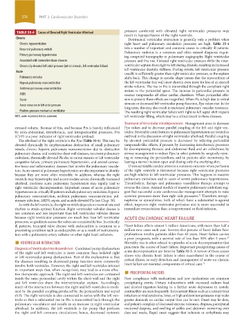
TABLE 35-4 Causes of Elevated Right Ventricular Afterload
result in hypoperfusion of the right ventricle.
Chronic Detrimental ventricular interaction is generally only a problem when
Chronic hypoventilation right heart and pulmonary circulation pressures are high. Table 35-4
Recurrent pulmonary emboli lists a number of important and common causes in critically ill patients.
Pulmonary embolus is a common and often missed diagnosis requir-
Primary pulmonary hypertension ing computed tomography or pulmonary angiography. Right ventricular
Associated with connective tissue diseases pressure and Pra rise. Elevated right ventricular pressure shifts the inter-
Chronically elevated left atrial pressure (mitral stenosis, left ventricular failure) ventricular septum from right to left during diastole, resulting in increased
left ventricular diastolic stiffness. During systole, left ventricular pressure
Acute
usually is sufficiently greater than right ventricular pressure, so the septum
Pulmonary embolus shifts back. This change in systolic shape means that the myocardium of
Hypoxic pulmonary vasoconstriction the left ventricular free wall must shorten even more for less of an ejected
stroke volume. The rise in Pra is transmitted through the compliant right
Acidemic pulmonary vasoconstriction
atrium to the pericardial space. The increase in pericardial pressure in
ARDS essence tamponades all other cardiac chambers. When pericardial effu-
Sepsis sion is present, these effects are magnified. When Pla is high due to mitral
stenosis or decreased left ventricular pump function, Ppa values rise. In the
Acute elevation in left atrial pressure
long term, this may also result in increased pulmonary vascular resistance.
Positive-pressure mechanical ventilation The resulting right ventricular failure with right-to-left septal shift impairs
ARDS, acute respiratory distress syndrome. left ventricular filling, which may be a critical insult in these diseases.
Treatment of Ventricular Interdependence: Management aims to decrease
stressed volume. Because of this, and because Pra is heavily influenced Ppa values and to decrease parallel coupling of the left and right ven-
by intra-abdominal, intrathoracic, and intrapericardial pressures, Pra tricles. Reversible contributions to pulmonary hypertension are treated as
(CVP) is a poor indicator of right ventricular preload. outlined in the discussion of right ventricular afterload. Parallel coupling
The afterload of the right ventricle is the Ppa (Table 35-4). This may be by elevated pericardial pressure is decreased by relieving pericardial
elevated chronically by emphysematous destruction of small pulmonary tamponade-like effects, if present; by decreasing intrathoracic pressures
vessels, chronic hypoxic pulmonary vasoconstriction due to obstructive by decompressing thoracic and abdominal fluid and air collections; by
pulmonary disease and restrictive chest wall diseases, recurrent pulmonary airway management to reduce Ppa; in select patients by surgically open-
embolism, chronically elevated Pla due to mitral stenosis or left ventricular ing or removing the pericardium; and in patients after sternotomy, by
congestive failure, primary pulmonary hypertension, and several connec- leaving a sternal incision open and closing only the overlying skin.
tive tissue and inflammatory diseases that involve the pulmonary vascula- Unresuscitatable cardiac arrest is a common outcome when perfusion
ture. Acute causes of pulmonary hypertension are also important to identify of the right ventricle is threatened because right ventricular pressures
because they are more often reversible. In addition, whereas the right are high relative to left ventricular pressures. This happens in massive
ventricle may hypertrophy and accommodate severe chronically increased pulmonary embolism and in cases of severe pulmonary hypertension.
afterload, moderate acute pulmonary hypertension may rapidly lead to Thrombolytic therapy and pulmonary vasodilator therapy attempt to
right ventricular decompensation. Important causes of acute pulmonary reverse the cause. Animal models of massive pulmonary embolism sug-
hypertension in critically ill patients include pulmonary embolism, hypoxic gest that successful acute cardiovascular management attempts to raise
pulmonary vasoconstriction, acidemic pulmonary vasoconstriction, pul- systemic pressures more than right-side pressures. Therefore, norepi-
71
monary infection, ARDS, sepsis, and acutely elevated Pla (see Chap. 38). nephrine or epinephrine, both of which have a substantial α-agonist
As with the left ventricle, the right ventricle depends on normal rate and effect, improves right ventricular perfusion and is more successful in
rhythm to attain optimal function. Right ventricular valvular disease is immediate resuscitation than is isoproterenol or fluid infusion.
less common and less important than left ventricular valvular disease
because right ventricular pressures are much less than left ventricular ACUTE ON CHRONIC HEART FAILURE
pressures, so gradients across the valves are considerably less. In critically
ill patients, tricuspid valve disease with endocarditis is common as a Heart failure affects almost 5 million Americans, with more than half a
preexisting condition such as endocarditis or as a result of instrumenta- million new cases each year. Seventy-five percent of heart failure hos-
tion with a pulmonary artery catheter or other right heart catheters. pitalizations involve patients older than 65 years. Heart failure carries
■ VENTRICULAR INTERACTION a poor prognosis, with a survival rate of less than 50% after 5 years.
27
Mortality rate is often related to episodes of acute decompensation that
Diagnosis of Ventricular Interdependence: Combined pump dysfunction punctuate the course of heart failure. Important precipitating causes of
of the right and left ventricles is more common than isolated right acute decompensation are listed in Table 35-5. A review of these causes
or left ventricular pump dysfunction. Part of the explanation is that shows why chronic heart failure is often exacerbated in the course of
the diseases resulting in decreased pump function more commonly critical illness, so early detection and management of acute-on-chronic
involve both ventricles. However, the right and left ventricles interact heart failure are essential components of critical care. 72
tive therapeutic approach. The right and left ventricles are contained ■ PRECIPITATING FACTORS
in important ways that, when recognized, may lead to a more effec-
inside the same pericardial cavity within the chest wall and the right Poor compliance with medications and new medications are common
and left ventricles share the interventricular septum. Accordingly, precipitating events. Dietary indiscretions with increased sodium load
much of the interaction between the right and left ventricles is medi- and alcohol ingestion leading to a further acute depression in systolic
ated by the parallel coupling produced by the pericardium and septal contractility are seen frequently. Intercurrent illness such as a urinary tract
shift. The right ventricle is also connected in series with the left ven- infection or viral syndrome, fever, or high ambient temperatures may make
tricle so that a substantial rise in Pla is transmitted back through the greater demands on cardiac output than can be met. Onset may be slow,
pulmonary vasculature and results in an increase in right ventricular and patients complain of decreased exercise tolerance, dyspnea, paroxysmal
afterload. In addition, the left ventricle is the pump that perfuses nocturnal dyspnea, and swelling of ankles and abdomen worsening over
the right and left coronary circulations; hence, decreased systemic days and weeks. Rapid onset suggests that ischemia or arrhythmia may
section03.indd 276 1/23/2015 2:07:06 PM