
Page 409 - Hall et al (2015) Principles of Critical Care-McGraw-Hill
P. 409
CHAPTER 36: Cardiac Arrhythmias, Pacing, Cardioversion, and Defibrillation in the Critical Care Setting 279
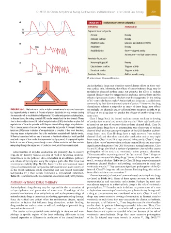
A TABLE 36-1 Mechanisms of Common Tachycardias
Arrhythmia Mechanism(s)
TP Supraventricular Tachycardia
RP
AV node Reentry
Accessory pathway Reentry
B Atrial tachycardia Enhanced automaticity or reentry
EAD Atrial flutter Reentry
Atrial fibrillation Onset—triggered activity
Maintenance—multiple wavelet reentry
Ventricular Tachycardia
C Monomorphic post MI Reentry
Catecholamine sensitive Triggered activity
Torsade de pointes Triggered activity
DAD Ventricular fibrillation Multiple wavelet reentry
AV, atrioventricular; MI, myocardial infarction.
D
Antiarrhythmic drugs may therefore exert different effects on these vari-
ous cardiac cells. Moreover, the effects of antiarrhythmic drugs may be
modified in diseased cardiac tissue. For example, the effects of sodium
channel blockers may be exaggerated in ischemic myocardium and the
effects of potassium channel blockers may be exaggerated in the setting
of left ventricular hypertrophy. Antiarrhythmic drugs are classified most
4
commonly by their dominant mechanism of action. However, this drug
4-6
classification scheme is imperfect as many of these drugs have effects
FIGURE 36-1. Mechanisms of cardiac arrhythmias—enhanced or abnormal automatic- on multiple ion channels and/or cell membrane receptors (Table 36-2).
ity, triggered activity or reentry. A. The rate of phase 4 depolarization may increase causing Efficacy of one drug does not predict the efficacy of another drug in the
the myocardial cell to reach the threshold potential (TP) earlier and spontaneously depolarize. same class.
In diseased tissue, the resting potential (RP) may be elevated and the time to reach TP may Class I drugs block the inward sodium current resulting in slowing
then be shortened (not shown). B. Early afterdepolarizations (EADs) develop late on phase 3 of of conduction in atrial and ventricular muscle. Their subclassification
6
repolarization of the action potential and if they reach threshold may trigger a depolarization. is based on the rate of recovery from sodium channel block. Class IA
This is the mechanism of torsade de pointes ventricular tachycardia. C. Delayed afterdepo- antiarrhythmic drugs have an intermediate rate of recovery of sodium
larizations (DADs) occur in diastole after repolarization is complete. If they reach threshold, channel block and may cause prolongation of the QRS duration at physi-
they may trigger a depolarization. This is the mechanism associated with digitalis toxicity. ologic heart rates. Class IB drugs have a rapid recovery from sodium
D. Reentry is associated with an area of anatomic or functional unidirection block (speckled channel block and thus slow ventricular conduction only at very rapid
area) and a region of slow conduction (represented by zigzag arrow). A propagated impulse heart rates. Class IA and IB drugs are used infrequently. Class IC drugs
blocks in one area of tissue, passes through an area of slow conduction and then conducts have a slow rate of recovery from sodium channel block and may cause
retrogradely through the original area of conduction block, which has now repolarized. significant prolongation of the QRS duration at resting heart rates. Class
IA and IC drugs also block a variety of potassium channels that causes
Abnormalities of impulse conduction are primarily due to reentry prolongation of the atrial and ventricular action potential durations.
(Fig. 36-1). Reentry requires an area of fixed or functional unidirec- This may manifest as prolongation of the Q ˙ t interval. Class II drugs are
2
tional block in one pathway, slow conduction in an alternate pathway β-adrenergic receptor blocking drugs. Some of these agents are selec-
7
and return of the impulse along the original path after this tissue has tive β -receptor blockers (Table 36-2). Class III drugs are predominantly
1
recovered excitability (Fig. 36-1D). Reentry is the mechanism of many potassium channel blockers and prolong repolarization in atrial and
8,9
types of supraventricular tachycardias (atrioventricular node reentry or ventricular muscle. They can cause significant prolongation of the Q ˙ t
in the Wolff-Parkinson-White syndrome) and scar-related ventricular interval. Class IV drugs are calcium channel blocking drugs that reduce
tachycardia (V ) that occurs following a myocardial infarction. intracellular calcium concentrations. 10
t
Table 36-1 summarizes the mechanisms of common tachyarrhythmias. The mechanism(s) of action of commonly used antiarrhythmic drugs
are listed in Table 36-2. Many of these agents are used to treat both
ANTIARRHYTHMIC DRUG THERAPY supraventricular and ventricular arrhythmias. Perhaps the most seri-
ous adverse response to antiarrhythmic drugs is the risk of ventricular
Antiarrhythmic drug therapy may be required for the termination of proarrhythmia. Proarrhythmia is defined as provocation of a new
7,11
tachyarrhythmias and prevention of recurrence. Knowledge of the arrhythmia or worsening of an existing arrhythmia during therapy with
potential mechanisms of an arrhythmia as well as the pharmacology of a drug at concentrations not considered to be toxic. Class IA and IC
antiarrhythmic drugs is important in selecting appropriate drug therapy. drugs may cause excessive slowing of conduction in diseased atrial or
Since the critical care patient often has multisystem disease, special ventricular muscle tissue that may exacerbate the clinical arrhythmia,
attention to factors that influence drug absorption, protein binding, for example, atrial flutter or V . These drugs increase the risk of sudden
t
drug metabolism and excretion as well as knowledge of potential drug cardiac death in patients following myocardial infarction. Thus, Class
interactions is essential. 4 I antiarrhythmic drugs are contraindicated in patients with ischemic
The cardiac action potential varies strikingly in duration and mor- heart disease and a prior myocardial infarction because of the risk of
phology in specific regions of the heart reflecting differences in ion ventricular proarrhythmia. Drugs that cause excessive prolongation
channel expression or differences in modulators of ion channel function. of the Q ˙ t interval may cause torsade de pointes V (Fig. 36-2). 4,7,11
4
t
section03.indd 279 1/23/2015 2:07:07 PM