
Page 470 - Hall et al (2015) Principles of Critical Care-McGraw-Hill
P. 470
340 PART 3: Cardiovascular Disorders
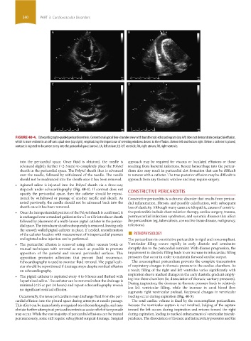
FIGURE 40-4. Echocardiography-guided pericardiocentesis. Conventional apical four-chamber view with transthoracic echocardiogram (top left) does not demonstrate pericardial effusion,
which is more evident in an off-axis apical view (top right), emphasizing the importance of selecting windows closest to the effusion. Bottom left and bottom right. Before a catheter is placed,
contrast is injected to document entry into the pericardial space (arrow). LA, left atrium; LV, left ventricle; RA, right atrium; RV, right ventricle.
into the pericardial space. Once fluid is obtained, the needle is approach may be required for viscous or loculated effusions or those
advanced slightly further (~2-3 mm) to completely place the Polytef resulting from bacterial infections. Recent hemorrhage into the pericar-
sheath in the pericardial space. The Polytef sheath then is advanced dium also may result in pericardial clot formation that can be difficult
over the needle, followed by withdrawal of the needle. The needle to remove with a catheter. The true posterior effusion may be difficult to
should not be readvanced into the sheath once it has been removed. approach from any thoracic window and may require surgery.
• Agitated saline is injected into the Polytef sheath via a three-way
stopcock under echocardiography (Fig. 40-4). If contrast does not
opacify the pericardial space, then the catheter should be reposi- CONSTRICTIVE PERICARDITIS
tioned by withdrawal or passage of another needle and sheath. As Constrictive pericarditis is a chronic disorder that results from pericar-
noted previously, the needle should not be advanced back into the dial inflammation, fibrosis, and possibly calcification, with subsequent
sheath once it has been removed. loss of elasticity. Although many cases are idiopathic, causes of constric-
• Once the intrapericardial position of the Polytef sheath is confirmed, it tive pericarditis include chest radiation therapy, cardiac surgery, trauma,
is exchanged over a standard guidewire for a 5 or 6 Fr introducer sheath postmyocardial infarction syndromes, and systemic diseases that affect
followed by placement of a multi-lumen pigtail catheter in the pericar- the pericardium (eg, tuberculosis, connective tissue disease, malignancy,
dial space. The introducer sheath subsequently is removed, leaving only infections).
of the catheter location with measurement of intrapericardial pressure ■ PATHOPHYSIOLOGY
the smooth walled pigtail catheter in place. If needed, reconfirmation
and agitated saline injection can be performed. The pericardium in constrictive pericarditis is rigid and noncompliant.
• The pericardial effusion is removed using either vacuum bottle or Ventricular filling occurs rapidly in early diastole and terminates
manual techniques with removal as much as possible to promote abruptly due to the pericardial restraint. With disease progression, the
apposition of the parietal and visceral pericardial surfaces. This impairment in diastolic filling leads to an increase in intracardiac filling
apposition promotes adhesions that prevent fluid recurrence. pressures that occur in order to maintain forward cardiac output.
Echocardiography is used to monitor fluid removal. The pigtail cath- The noncompliant pericardium prevents the complete transmission
eter should be repositioned if drainage stops despite residual effusion of respiratory changes in thoracic pressure to the cardiac chambers. As
on echocardiography. a result, filling of the right and left ventricles varies significantly with
• The pigtail catheter is aspirated every 4 to 6 hours and flushed with respiration due to marked changes in the early diastolic gradient empty-
heparinized saline. The catheter can be removed when the drainage is ing into these chambers (ie, dissociation of thoracic-cavitary pressures).
minimal (<25 cc per 24 hours) and repeat echocardiography reveals During inspiration, the decrease in thoracic pressure leads to relatively
no significant residual effusion. less left ventricular filling, while the increase in caval blood flow
augments right ventricular preload. Reciprocal changes in ventricular
Occasionally, the tense pericardium may discharge fluid from the peri- loading occur during expiration (Fig. 40-5).
cardial effusion into the pleural space during attempts at needle passage. The total cardiac volume is fixed by the noncompliant pericardium.
This effect can be immediately recognized on echocardiography, and may Because the ventricular septum is not involved, bulging of the septum
obviate further attempts at pericardiocentesis as acute relief of tamponade toward the left occurs during inspiration and returns toward the right
may occur. While the vast majority of pericardial effusions can be treated during expiration, leading to marked enhancement of ventricular interde-
percutaneously, some still require subxyphoid surgical drainage. Surgical pendence. The dissociation of thoracic and intracavitary pressures and the
section03.indd 340 1/23/2015 2:07:43 PM