
Page 474 - Hall et al (2015) Principles of Critical Care-McGraw-Hill
P. 474
344 PART 3: Cardiovascular Disorders
diagnostic ability, it has largely replaced catheterization as a diagnostic rheumatic AS, stenosis is caused by fusion of the commissures with
modality in valvular disease; the latter is usually indicated only when scarring and eventual calcification. It is less commonly seen in the
discrepancies between echocardiographic and clinical findings are Western world and is invariably accompanied by various degrees of
noted. Other routine evaluations (electrocardiography, chest x-ray) mitral valve disease.
are of obvious utility in critically ill patients. In complex situations, ■
cardiac CT and MRI can further complete the diagnosis, but require NATURAL HISTORY
transportation of the critically ill patient to the specific areas, which is Classically, AS begins with a prolonged asymptomatic period, in which
cumbersome. morbidity and mortality are very low. However, once even moderate dis-
In this chapter, we will further review the etiology, pathophysiology, ease is present, AS is a relentlessly progressive disease. The average rate of
clinical presentation, diagnostic evaluation, and management of critical progression is an increase in mean pressure gradient of 7 mm Hg per year,
illness in the context of major valvular disease. Acute prosthetic valvular and a decrease in valve area of 0.1 cm per year, but there is marked indi-
5
2
disease and infective endocarditis will be presented at the end of the vidual variability. Regular clinical follow-up is mandatory in all patients
chapter. with asymptomatic mild to moderate AS.
AORTIC STENOSIS ■ PATHOPHYSIOLOGY
■ ETIOLOGY The key hemodynamic change in AS is a progressively increasing
resistance to blood flow. In the Gorlin equation, the cardiac output
6
The prevalence of significant aortic valvular heart disease (moderate is directly proportional to the square root of the pressure gradient.
severity or worse) increases with age, occurring in only 0.7% of those Therefore, small changes in both cardiac output and valve area may have
age 18 to 44 years but in 13.3% of adults 75 years and older. Native aor- significant effects on the pressure gradient. Such large variations may
1
tic valve stenosis is the most common valvular lesion in clinical prac- lead to confusion in classification of disease severity, as currently used
tice, followed by mitral regurgitation (25%), and multivalve disease criteria to define severe AS are not necessarily simultaneously present
2
(20%). According to location, aortic stenosis (AS) can be classified in all patients. Intuitively, aortic valve area should be the best estimate
as subvalvular, valvular, or supravalvular (Fig. 41-1). Subvalvular of AS severity as it represents the anatomical obstacle to left ventricular
and supravalvular stenoses result from focal (isolated membrane) or outflow, and is less prone to variations under hemodynamic conditions.
2
extended (tubular) stenotic lesions. Regardless of lesion type, they lead Indeed, a valve area of less than 1.0 cm was associated with unfavorable
to impaired flow in the left ventricular outflow tract or aortic root, and are outcome regardless of gradient or symptoms. 7
indistinguishable from valvular AS from a hemodynamic perspective. Stroke volume and cardiac output are initially maintained by
Therefore, their management in critically ill patients is similar to val- hypertrophy of the left ventricle. The increased wall thickness leads to
vular AS. maintaining wall stress within normal limits, and explains why cardiac
The most common cause of valvular AS is degenerative disease of a output, ejection fraction, and left ventricular cavity dimensions are
tricuspid aortic valve. This is very common after the age of 70, leading maintained for a long period. Once compensatory mechanisms are
to significant morbidity and mortality. Surgical series have reported overwhelmed by progressive stenosis, the cardiac output declines, and
the incidence of degenerative AS as high as 10% to 30% but the true the left ventricle eventually enlarges. Note that transvalvular gradient
3
prevalence is likely underestimated considering that many patients actually declines in these late stages, leading to the “low cardiac output,
8
are not referred for surgical correction. Degeneration of a congenitally low gradient” type AS. Classically, only patients with low EF were
malformed aortic valve (bicuspid or unicuspid aortic valves) occurs included in this category. More recently, emphasis has been placed
earlier in life, and patients present with significant valvular disease in on patients with pseudonormal left ventricular function. These are
the middle or late adult life. It is estimated that 1% to 2% of aortic valves individuals in whom a low transvalvular gradient is present despite
are bicuspid, making this one of the most common congenital heart preserved EF; the low cardiac output in this situation is explained by
malformations. In both tricuspid and congenitally malformed valves, a combination of low stroke volume and increased valvuloarterial
4
9
degeneration of the valve progresses from the base of the cusps to the impedance. In the absence of surgical intervention the outcome of
leaflets, eventually causing a reduction in leaflet motion and effective patients with low cardiac output and severe AS is poor, regardless
valve area; commissural fusion is a late phenomenon. Calcific AS is of type (low EF or normal EF).
an active disease process characterized by lipid accumulation, inflam- Left ventricular hypertrophy maintains contractile function, but
mation, and calcification, with many similarities to atherosclerosis. In will ultimately lead to relaxation abnormalities (diastolic dysfunction),
resulting in elevated left atrial pressure and secondary pulmonary
hypertension. Ischemia may develop due to both concomitant coro-
nary artery disease (present in ∼50% of patients with severe AS) and
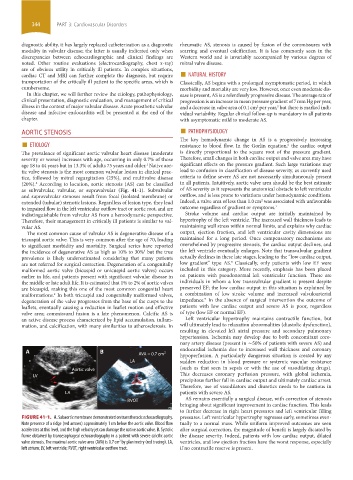
A B AVA = 0.7 cm 2 endocardial ischemia due to increased wall thickness and coronary
hypoperfusion. A particularly dangerous situation is created by any
LA sudden reduction in blood pressure or systemic vascular resistance
LA
(such as that seen in sepsis or with the use of vasodilating drugs).
Aortic valve
LV This decreases coronary perfusion pressure, with global ischemia,
precipitous further fall in cardiac output and ultimately cardiac arrest.
Therefore, use of vasodilators and diuretics needs to be cautious in
LA patients with severe AS.
RVOT AS remains essentially a surgical disease, with correction of stenosis
bringing about significant improvement in cardiac function. This leads
to further decrease in right heart pressures and left ventricular filling
FIGURE 41-1. A. Subaortic membrane demonstrated on transthoracic echocardiography. pressures. Left ventricular hypertrophy regresses early, sometimes even-
Note presence of a ridge (red arrows) approximately 1 cm below the aortic valve. Blood flow tually to a normal mass. While uniform improved outcomes are seen
accelerates at this level, and the high velocity jet can damage the native aortic valve. B. Systolic after surgical correction, the magnitude of benefit is largely dictated by
frame obtained by transesophageal echocardiography in a patient with severe calcific aortic the disease severity. Indeed, patients with low cardiac output, dilated
valve stenosis. The maximal aortic valve area (AVA) is 0.7 cm by planimetry (red tracing). LA, ventricles, and low ejection fraction have the worst response, especially
2
left atrium; LV, left ventricle; RVOT, right ventricular outflow tract. if no contractile reserve is present.
section03.indd 344 1/23/2015 2:07:46 PM