
Page 472 - Hall et al (2015) Principles of Critical Care-McGraw-Hill
P. 472
342 PART 3: Cardiovascular Disorders
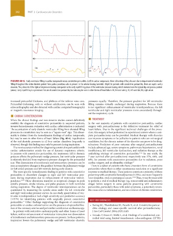
80 80
FA
LV
*
40 40
a v
RA a v x
x
RA y
FIGURE 40-6. Early ventricular filling in cardiac tamponade versus constrictive pericarditis. (Left) In cardiac tamponade, there is blunting of the y descent due to impairment of ventricular
filling throughout the entire diastolic period. Note pulsus paradoxus also is present in the arterial tracing (asterisk). (Right) In patients with constrictive pericarditis, there are rapid x and y
descents. The y descent of the right atrial pressure tracing corresponds to the early rapid filling phase of the ventricular pressure tracing, which demonstrates the typical dip and plateau pattern
(arrow). Early rapid filling is a prominent feature of constrictive pericarditis, but also may be seen in other forms of heart failure. FA, femoral artery; LV, left ventricle; RA, right atrial.
increased pericardial thickness, and plethora of the inferior vena cava. pressures equally. Therefore, the pressure gradient for left ventricular
Pericardial thickening, with or without calcification, can be seen with filling remains virtually unchanged during respiration. Because there
echocardiography and also detected with cardiac computed tomography is not significant enhancement of ventricular interdependence, the left
or magnetic resonance imaging. ventricular and right ventricular pressures move concordantly through-
■
out the respiratory cycle.
■
When the clinical findings and non-invasive studies cannot definitively TREATMENT
CARDIAC CATHETERIZATION
establish the diagnosis of constrictive pericarditis in suspected patients, In the vast majority of patients with constrictive pericarditis, cardiac
invasive hemodynamic evaluation with cardiac catheterization is indicated. surgery with pericardiectomy is the definitive treatment for relief of
The accentuation of early diastolic ventricular filling from elevated filling heart failure. Due to the significant technical challenges of the proce-
pressures in constriction may be seen as a “square-root” sign. This abnor- dure, this surgery is best performed in experienced centers where a com-
mality is distinct from the hemodynamic findings of cardiac tamponade, plete pericardiectomy can be provided. Medical therapy with diuretics
but may be seen in other forms of heart failure (Fig. 40-6). Equalization can improve symptoms or be palliative in patients who are not surgical
of the end-diastolic pressures in all four cardiac chambers frequently is candidates, but the chronic nature of the disorder can prove to be drug-
observed, though this finding may only be present during inspiration. refractory. Predictors of poor outcome after surgical pericardiectomy
The most accurate method for diagnosing constrictive pericarditis with include advanced age, severe symptoms, pulmonary hypertension, renal
cardiac catheterization entails the use of dynamic respiratory criteria. insufficiency, left ventricular dysfunction, and radiation therapy as the
In patients with constrictive pericarditis, the inspiratory fall in thoracic underlying etiology of constrictive pericarditis. 15,16 In one study, the
pressure affects the pulmonary wedge pressure, but ventricular pressure 7-year survival after pericardiectomy respectively was 27%, 66%, and
is relatively shielded from respiratory pressure changes by the pericardial 88%, for patients with constrictive pericarditis due to radiation, prior
scar. This dissociation of intrathoracic and intracavitary pressures can be cardiac surgery, and an idiopathic etiology. 17
seen as respiratory changes in the gradient between the pulmonary wedge There is a subset of patients who have a transient form of constrictive
(or left atrial) pressure and left ventricle during early diastole. pericarditis where there is either spontaneous resolution or a significant
The most specific hemodynamic finding in patients with constrictive response to medical therapy. These patients constitute a minority of those
pericarditis is discordant changes in right and left ventricular pres- presenting with constrictive hemodynamics (<25%), and more frequently
sures during respiration due to enhancement of ventricular interde- have idiopathic, viral, or postsurgical causes. 18-20 Thus, it may be reasonable
pendence. 12,13 These alterations manifest as reciprocal changes in peak to perform a trial of medical therapy (eg, nonsteroidal anti-inflammatory
systolic pressure, stroke volume, and pulse pressure in both ventricles drugs) before surgery in some patients presenting with constrictive
during respiration. The degree of ventricular interdependence can be pericarditis, particularly those with mild symptoms, a potentially revers-
quantitated by measuring the systolic areas under the left ventricular ible cause of acute inflammation, and no evidence of chronic constriction.
and right ventricular pressure curves. In one study, quantitation of ven-
tricular interdependence had a high sensitivity and predictive accuracy
(>97%) for identifying patients with surgically proven constrictive KEY REFERENCES
pericarditis. Other findings supporting the diagnosis of constrictive
14
pericarditis at cardiac catheterization are the presence of epicardial fixa- • Bertog SC, Thambidorai SK, Parakh K, et al. Constrictive pericar-
tion of the coronary arteries and pericardial calcification on fluoroscopy. ditis: etiology and cause-specific survival after pericardiectomy.
In patients with restrictive cardiomyopathy and other forms of heart J Am Coll Cardiol. 2004;43(8):1445.
failure, neither enhancement of ventricular interaction nor dissociation • Ferrada P, Evans D, Wolfe L, et al. Findings of a randomized con-
of intrathoracic and intracavitary pressures are present. In these patients, trolled trial using limited transthoracic echocardiogram (LTTE)
inspiration lowers the pulmonary wedge and left ventricular diastolic
section03.indd 342 1/23/2015 2:07:45 PM