
Page 475 - Hall et al (2015) Principles of Critical Care-McGraw-Hill
P. 475
CHAPTER 41: Valvular Heart Disease 345
■ CLINICAL PRESENTATION comprehensive interrogation of the aortic valve gradient from multiple
In the critically ill patient, presentation with severe AS is a reflection of windows must be performed to ensure capturing the highest gradient.
In the current ACC/AHA Valvular Heart Disease Guidelines, AS
5
acute decompensation, often due to a concurrent condition because AS
severity progresses otherwise slowly. Acute decompensated heart failure is classified into mild, moderate, and severe according to echocardio-
graphic findings. A velocity >4 m/s, gradient >40 mm Hg, and valve
in patients with severe AS is characterized by presence of dyspnea, less 2
so by angina or syncope. Some patients complain of nonspecific symp- area <1.0 cm are consistent with severe disease. These criteria have
been criticized as being intrinsically discordant, as up to 30% of
11
toms such as fatigue, dizziness, or palpitations. A particular presenta- 2
tion is that of decompensated heart failure in patients with severe AS patients with calculated valve area of less than 1 cm will not have veloci-
ties and gradients in the severe range. Some of these patients have low
undergoing noncardiac surgery. In this situation, large volume shifts
and vasodilation associated with surgical procedure and anesthesia may gradients due to low cardiac output and true AS, while others may have
low calculated valve areas in the context of a nonvalvular myopathic
lead to acute decompensation. Indeed, among valvular diseases AS is
associated with the highest risk of perioperative complications, up to process rendering the left ventricle unable to generate enough pres-
sure for full valve opening (pseudosevere AS). Low-dose dobutamine
10% mortality in some series. 10 12
Physical examination can suggest presence of AS. The apical impulse echocardiography is helpful in diagnosis. Indeed, when cardiac output
is usually sustained, and is not significantly displaced unless the ven- increases on the background of true severe AS, this will result in a cor-
tricle has dilated. The hallmark auscultatory findings are presence of the responding increase in transvalvular gradients, and the calculated valve
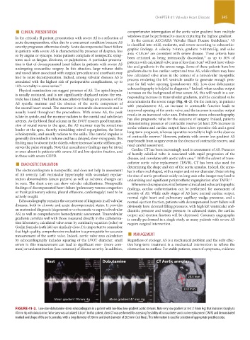
area remains in the severe range (Fig. 41-2). On the contrary, in patients
AS systolic murmur and the absence of the aortic component of
the second heart sound. The murmur is crescendo-decrescendo and is with pseudosevere AS, an increase in contractile function leads to
improved opening of the aortic valve, and the increase in cardiac output
usually heard throughout the precordium. With severe AS, the peak
is late in systole, and the murmur radiates to the carotid and subclavian results in an increased valve area. Dobutamine stress echocardiography
has also prognostic value for the outcome of surgery. Indeed, patients
arteries. As the blood fluid column in the LVOT ensures good transmis-
sion of sound waves to the apex, the AS murmur may be sometimes demonstrating presence of contractile reserve (at least 20% increase in
stroke volume and cardiac output) have a low operative risk and a good
louder at the apex, thereby mimicking mitral regurgitation; the latter
is holosystolic, and usually radiates to the axilla. The carotid impulse is long-term prognosis, whereas operative mortality is high in the absence
of contractile reserve. However, patients with severe low-gradient AS
8
generally diminished in volume and has a delayed, slow-rising peak; this
finding may be absent in the elderly, where increased aortic stiffness pre- may benefit from surgery even in the absence of contractile reserve, and
need careful assessment.
serves the pulse strength. Note that auscultatory findings may be trivial
Cardiac CT has been increasingly used in assessment of AS. Presence
or even absent in patients with severe AS and low ejection fraction, and of heavily calcified valve is associated with rapid progression of the
in those with severe COPD. disease, and correlates with aortic valve area. With the advent of trans-
■ DIAGNOSTIC EVALUATION catheter aortic valve replacement (TAVR), CT has been also used for
13
The electrocardiogram is nonspecific, and does not help in assessment determining the shape and size of the aortic annulus. Indeed, the annu-
lus is often oval shaped, with a major and minor diameter. Determining
of AS severity. Left ventricular hypertrophy with secondary repolar- the size of aortic prosthesis solely on long-axis echo images may lead to
ization abnormalities (strain pattern) as well as ischemic changes can undersizing and significant periprosthetic regurgitation after TAVR. 14
be seen. The chest x-ray can show valvular calcifications. Nonspecific Whenever discrepancies exist between clinical and echocardiographic
findings of decompensated heart failure (pulmonary venous congestion findings, cardiac catheterization can be performed for assessment of
or frank pulmonary edema, pleural effusions, cardiomegaly) need to be severity of AS. While early stages of AS have normal cardiac output,
actively sought. normal right heart and pulmonary capillary wedge pressures, and a
Echocardiography remains the cornerstone of diagnosis in all valvular normal ejection fraction, patients with decompensated heart failure will
diseases, both in chronic and acute decompensated states. It provides obviously have elevated filling pressures, with high left ventricular end-
an anatomical diagnosis (degenerative vs rheumatic vs supra/subvalvular diastolic pressure and wedge pressure. In advanced states, the cardiac
AS) as well as comprehensive hemodynamic assessment. Transvalvular output and ejection fraction will be depressed. Coronary angiography
gradients correlate well with those measured directly in the catheteriza- is usually performed in a single study, as many patients with severe AS
tion laboratory; calculated valve areas by continuity equation (echo) or require surgical intervention.
Gorlin formula (cath lab) are similarly close. It is important to remember
measurement of the aortic valve. Indeed, aortic valve area calculation ■ MANAGEMENT
that high-quality, comprehensive evaluation is a prerequisite for accurate
by echocardiography includes squaring of the LVOT diameter; small Regardless of etiology, AS is a mechanical problem and the only effec-
errors in this measurement can lead to significant over- (more com- tive long-term treatment is a mechanical intervention to relieve the
mon) or underestimation (less common) of disease severity. In addition, obstruction to outflow. For stable patients, onset of symptoms, evidence
Rest Dobutamine CT Aortic annulus
20.5 mm
30.0 mm
Mean gradient 19 mm Hg Mean gradient 47 mm Hg
FIGURE 41-2. Low-dose dobutamine-stress echocardiogram in a patient with low-flow, low-gradient aortic stenosis. Note very low gradient at rest (19 mm Hg) that increases sharply to
47 mm Hg with dobutamine. Valve area was calculated 0.8 cm . In this patient, chest CT was performed for assessing feasibility of transcatheter aortic valve replacement (TAVR) and demonstrated
2
marked oval shape of the aortic annulus, with a long diameter of 30 mm and short diameter of 20.5 mm (red lines). This information is used for selection of appropriate prosthesis size.
section03.indd 345 1/23/2015 2:07:48 PM