
Page 598 - Hall et al (2015) Principles of Critical Care-McGraw-Hill
P. 598
418 PART 4: Pulmonary Disorders
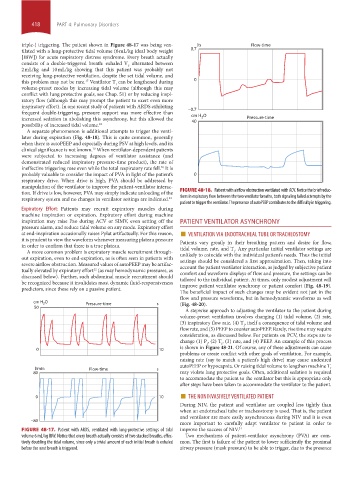
triple-) triggering. The patient shown in Figure 48-17 was being ven- l/s Flow-time
tilated with a lung-protective tidal volume (6 mL/kg ideal body weight 0.7
[IBW]) for acute respiratory distress syndrome. Every breath actually
consists of a double-triggered breath: exhaled V alternated between
T
2 mL/kg and 10 mL/kg showing that this patient was probably not
receiving lung-protective ventilation, despite the set tidal volume, and
17
this problem may not be rare. Ventilator T can be lengthened during 0
I
volume-preset modes by increasing tidal volume (although this may
conflict with lung protective goals, see Chap. 51) or by reducing inspi-
ratory flow (although this may prompt the patient to exert even more
inspiratory effort). In one recent study of patients with ARDS exhibiting
frequent double-triggering, pressure support was more effective than −0.7
2
increased sedation in abolishing this asynchrony, but this allowed the cm H O Pressure-time
40
possibility of increased tidal volume. 18
A separate phenomenon is additional attempts to trigger the venti-
lator during expiration (Fig. 48-18). This is quite common, generally
when there is autoPEEP and especially during PSV at high levels, and its
clinical significance is not known. When ventilator-dependent patients
19
were subjected to increasing degrees of ventilator assistance (and
demonstrated reduced inspiratory pressure-time product), the rate of
ineffective triggering rose even while the total respiratory rate fell. It is
20
probably valuable to consider the impact of PVA in light of the patient’s 0
respiratory drive. When drive is high, PVA should be addressed by
manipulation of the ventilator to improve the patient-ventilator interac-
tion. If drive is low, however, PVA may simply indicate unloading of the FIGURE 48-18. Patient with airflow obstruction ventilated with ACV. Notice the brief reduc-
respiratory system and no changes in ventilator settings are indicated. 21 tions in expiratory flow between the two ventilator breaths, both signaling failed attempts by the
patient to trigger the ventilator. The presence of autoPEEP contributes to the difficulty in triggering.
Expiratory Effort: Patients may recruit expiratory muscles during
machine inspiration or expiration. Expiratory effort during machine
inspiration may raise Pao during ACV or SIMV, even setting off the PATIENT VENTILATOR ASYNCHRONY
pressure alarm, and reduce tidal volume on any mode. Expiratory effort
at end-inspiration occasionally raises Pplat artifactually. For this reason, ■ VENTILATION VIA ENDOTRACHEAL TUBE OR TRACHEOSTOMY
it is prudent to view the waveform whenever measuring plateau pressure Patients vary greatly in their breathing pattern and desire for flow,
in order to confirm that there is a true plateau. tidal volume, rate, and T . Any particular initial ventilator settings are
A more common problem is expiratory muscle recruitment through- unlikely to coincide with the individual patient’s needs. Thus the initial
I
out expiration, even to end-expiration, as is often seen in patients with settings should be considered a first approximation. Then, taking into
severe airflow obstruction. Measured values of autoPEEP may be artifac- account the patient ventilator interaction, as judged by subjective patient
22
tually elevated by expiratory effort (as may hemodynamic pressures, as comfort and waveform displays of flow and pressure, the settings can be
discussed below). Further, such abdominal muscle recruitment should tailored to the individual patient. At times, only modest adjustment will
be recognized because it invalidates most dynamic fluid-responsiveness improve patient ventilator synchrony or patient comfort (Fig. 48-19).
predictors, since these rely on a passive patient.
The beneficial impact of such changes may be evident not just in the
flow and pressure waveforms, but in hemodynamic waveforms as well
cm H 2 O Pressure-time s (Fig. 48-20).
50
A stepwise approach to adjusting the ventilator to the patient during
volume-preset ventilation involves changing (1) tidal volume, (2) rate,
(3) inspiratory flow rate, (4) T , itself a consequence of tidal volume and
I
flow rate, and (5) PEEP to counter autoPEEP. Rarely, rise time may require
consideration, as discussed below. For patients on PCV, the steps are to
change (1) P , (2) T , (3) rate, and (4) PEEP. An example of this process
I
I
0 10 is shown in Figure 48-21. Of course, any of these adjustments can cause
problems or create conflict with other goals of ventilation. For example,
raising rate (say to match a patient’s high drive) may cause undesired
autoPEEP or hypocapnia. Or raising tidal volume to lengthen machine T
l/min Flow-time s I
80 may violate lung protective goals. Often, additional sedation is required
to accommodate the patient to the ventilator but this is appropriate only
after steps have been taken to accommodate the ventilator to the patient.
■
0 10 THE NONINVASIVELY VENTILATED PATIENT
During NIV, the patient and ventilator are coupled less tightly than
when an endotracheal tube or tracheostomy is used. That is, the patient
and ventilator are more easily asynchronous during NIV and it is even
−80
more important to carefully adapt ventilator to patient in order to
FIGURE 48-17. Patient with ARDS, ventilated with lung-protective settings of tidal improve the success of NIV. 21
volume 6 mL/kg IBW. Notice that every breath actually consists of two stacked breaths, effec- Two mechanisms of patient-ventilator asynchrony (PVA) are com-
tively doubling the tidal volume, since only a trivial amount of each initial breath is exhaled mon. The first is failure of the patient to lower sufficiently the proximal
before the next breath is triggered. airway pressure (mask pressure) to be able to trigger, due to the presence
section04.indd 418 1/23/2015 2:19:11 PM