
Page 654 - Hall et al (2015) Principles of Critical Care-McGraw-Hill
P. 654
CHAPTER 53: Extracorporeal Lung Support 473
membrane oxygenator to temporarily take over the functions of the lung. When severe ARDS patients are transferred to us for possible ECMO
While on ECMO, mechanical ventilator settings are adjusted to minimize evaluation, we place a right internal jugular and right femoral venous
VILI and to maximize the recruitment to functional residual capacity catheter in the event that ECMO is required, so that VV-ECMO can-
with an algorithm that aims to normalize body physiology and minimize nulation can proceed expeditiously. The ECMO charge specialist is
barotrauma. This algorithm used in 141 patients with respiratory failure contacted, and an ECMO circuit and ECMO blood pack is prepared.
referred for consideration of ECMO yielded a survival rate of 62% in ■
ratio of 66). 41 CANNULATION
patients with severe ARDS (median initial Pa O 2 /Fi O 2
The primary indication for use of ECMO in patients with severe The majority of patients with severe hypoxemia are managed with
respiratory failure is when the risk of dying from ARDS is considered VV-ECMO. Adult patients are typically cannulated percutaneously
greater than 80% despite optimal ventilator and medical management. with 21 to 23 French catheters for drainage and infusion of blood.
ratio of less than 70 on 100% oxygen. Percutaneous venous ECMO cannula insertion is the standard, but
This translates to a Pa O 2 /Fi O 2
surgical cutdown is required in some circumstances. Traditional can-
INITIATION OF ECMO nulation for VV-ECMO has been a two-cannula system with venous
drainage from the right femoral vein and return to the right atrium
Once the patient is considered an appropriate candidate for ECMO, the via a right internal jugular vein cannula (Fig. 53-3). A single bicaval
potential risks and complications associated with ECMO should be dis- dual lumen cannula placed in the internal jugular position is preferred
cussed with the patient’s legal surrogate, and written informed consent if able to be positioned appropriately, since early mobilization of the
should be obtained. We have a template ECMO consent form available ICU patient is then feasible. This ECMO cannula allows simultaneous
on our internal ECMO Web site that is printed in the ICU and then removal of blood from both the superior and inferior vena cavae with
scanned into the patient’s electronic medical record. return of blood into the right atrium with minimal recirculation.
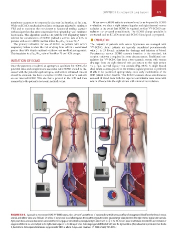
FIGURE 53-3. Approach to veno-venous ECMO (VV-ECMO) cannulation. Left panel shows the use of two cannulae with (1) venous outflow of deoxygenated blood from the femoral venous
cannula and inferior vena cava (IVC) and (2) inflow of oxygenated blood after it passes through the oxygenator where gas exchange takes place into the right internal jugular vein cannula.
Right panel shows a bicaval dual-lumen cannula in the internal jugular vein extending through the right atrium with its tip in the IVC. Venous blood is withdrawn from the IVC and reinfusion of
oxygenated blood is via a medial port in the right atrium adjacent to the tricuspid valve, delivering oxygenated blood directly into the right ventricle. (Reproduced with permission from Brodie
D, Bacchetta M. Extracorporeal membrane oxygenation for ARDS in adults. N Engl J Med. November 17, 2011;365(20):1905-1914.)
section04.indd 473 1/23/2015 2:19:54 PM