
Page 23 - The Netter Collection of Medical Illustrations - Integumentary System_ Volume 4 ( PDFDrive )
P. 23
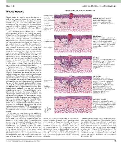
Plate 1-8                                                                             Anatomy, Physiology, and Embryology

                                                                          HEALING OF INCISED, SUTURED SKIN WOUND
        WOUND HEALING
                                                  Blood clot
        Wound healing is a complex process that involves an   Epithelium
        orderly  and  sequential  series  of  interactions  among                                         Immediately after incision
        multiple  cell  types  and  tissue  structures.  Classically,   Dermis                            Blood clot with fine fibrin
        wound  healing  has  been  divided  into  three  phases:   Incision                               network forms in wound.
        inflammation, new tissue formation, and matrix forma-                                             Epithelium thickens at wound
                                                                                                          edges.
        tion and remodeling. Each of these phases is unique,   Suture
        and particular cell types play key roles in the different   Subcutaneous
        phases.                                   fatty tissue
          Once a disruption of the skin barrier occurs, a cascade
        of  inflammatory  mediators  are  released,  and  wound
        healing begins. The disruption of dermal blood vessels
        allows extravasation of blood into the tissues. The rup-                                          24-48 hours
        tured  vessels  undergo  immediate  vasoconstriction.                                             Epithelium begins to grow down
                                                                                                          along cut edges and along suture
        Platelets begin the process of coagulation and initiate   Lymphocytes                             tract. Leukocyte infiltration,
        the  earliest  phase  of  inflammation.  The  formation  of                                       chiefly round cells (lymphocytes)
        the  earliest  blood  clot  provides  the  foundation  for                                        with few giant cells, occurs and
        future cell migration into the wound. Many inflamma-                                              removes bacteria and necrotic
        tory  mediators  are  released  during  this  initial  phase.   Giant cells                       tissue.
        Once initial homeostasis is achieved, the platelets dis-
        charge  the  contents  of  their  alpha  granules  into  the
        extravascular space. Alpha granules contain fibrinogen,
        fibronectin,  von  Willebrand’s  factor,  factor  VIII,  and
        many other proteins. The fibrinogen is converted into
        fibrin, which aids in formation of the fibrin clot. Plate-                                        5-8 days
        lets also play a critical role in releasing growth factors                                        Epithelial downgrowth advances.
        and  proteases.  The  best  known  of  these  is  platelet-                                       Fibroblasts grow in from deeper
        derived  growth  factor  (PDGF),  which  helps  mediate                                           tissues and add collagen
        the formation of the initial granulation tissue.  Fibroblasts                                     precursors and glycoproteins to
                                                                                                          matrix. Cellular infiltration
          During the late portion of the inflammatory phase,                                              progresses.
        leukocytes are seen for the first time. Neutrophils make
        up  the  largest  component  of  the  initial  leukocyte
        response.  Neutrophils  are  drawn  into  the  area  by
        various cytokines and adhere to the activated vascular
        endothelium. They enter the extravascular space by a                                              10-15 days
        process of diapedesis. These early-arriving neutrophils                                           Capillaries grow in from
        are  responsible  for  the  recruitment  of  more  neutro-  Keratinizing                          subcutaneous tissue, forming
        phils, and they also begin the process of killing bacteria   pearl                                granulation tissue. Epithelium
        by  use  of  their  internal  myeloperoxidase  system.                                            bridges incision; epithelial
        Through  the  production  of  free  radicals,  neutrophils                                        downgrowths regress, leaving
        are efficient at killing large numbers of bacteria. Neu-  Capillary                               keratinizing pearls behind.
        trophil  activity  continues  for  a  few  days,  unless  the   ingrowth                          Fibrosed clot (scab) is being
        wound is contaminated with bacteria. Once the neutro-                                             pushed out. Collagen formation
        phil  activity  has  cleared  the  wound  of  bacteria  and                                       progresses and cellular
                                                                                                          infiltration abates.
        other  foreign  particles,  monocytes  are  recruited  into
        the  wound  and  activated  into  macrophages.  Macro-
        phages are critical in clearing the wound of neutrophils
        and  any  remaining  cellular  and  bacterial  debris.                                            3 weeks–9 months
        Macrophages  are  capable  of  producing  nitrous  oxide,                                         Epithelium is thinned to near
        which  can  kill  bacteria  and  has  also  been  shown  to                                       normal. Tensile strength of tissue
        decrease  viral  replication.  Macrophages  also  release                                         is increased owing to production
        various cytokines, including PDGF, interleukin-6, and                                             and cross-linking of collagen
        granulocyte colony-stimulating factor (G-CSF), which                                              fibers; elastic fibers reappear
        in turn recruit more monocytes and fibroblasts into the                                           later.
        wound.
          At this point, new tissue formation, the proliferative
        phase of wound healing, has begun. This phase typically
        begins on the third day and ends about 14 days after the
        initial  insult.  It  is  marked  by  reepithelialization  and   contain the keratin pairs 5,14 and 6,16. They secrete   The final phase of wound healing involves scar matu-
        formation  of  granulation  tissue.  Reepithelialization   vascular  endothelial  growth  factor,  which  promotes     ration  and  tissue  remodeling.  This  phase  overlaps  in
        occurs  by  the  movement  of  epithelial  cells  (keratino-  the  production  of  dermal  blood  vessels.  At  the  same   time with the first two phases; it is said to begin with
        cytes) from the free edge of the wound slowly across the   time  the  keratinocytes  are  migrating,  the  underlying   the  production  of  the  first  granulation  tissue.  This
        wound  defect.  The  migrating  cells  have  the  distinct   fibroblasts  are  synthesizing  a  backbone  matrix,  made     phase extends for months and is complete when most
        phenotype  of  basal  keratinocytes.  It  is  believed  that     up  predominantly  of  type  III  collagen  and  some     of the collagen III and fibronectin have been replaced
        a  low  calcium  concentration  in  the  wound  causes     proteoglycans.  Some  of  the  fibroblasts  are  converted   by mature type I collagen. In the final mature scar, the
        the  keratinocytes  to  take  on  the  characteristics  of     into  myofibroblasts  by  PDGF  and  tumor  growth   collagen  fibers  are  oriented  in  large  bundles  running
        basal keratinocytes. PDGF is an important stimulant for   factor-β1. These myofibroblasts are important in that   perpendicular  to  the  basement  membrane  zone.  The
        keratinocytes and is partially responsible for this migra-  they cause the overlying wound to contract, decreasing   resulting scar has only 80% of the tensile strength of
        tion  across  the  wound.  The  migrating  keratinocytes   its surface.             the uninjured skin.


        THE NETTER COLLECTION OF MEDICAL ILLUSTRATIONS                                                                            9