
Page 236 - The Netter Collection of Medical Illustrations - Integumentary System_ Volume 4 ( PDFDrive )
P. 236
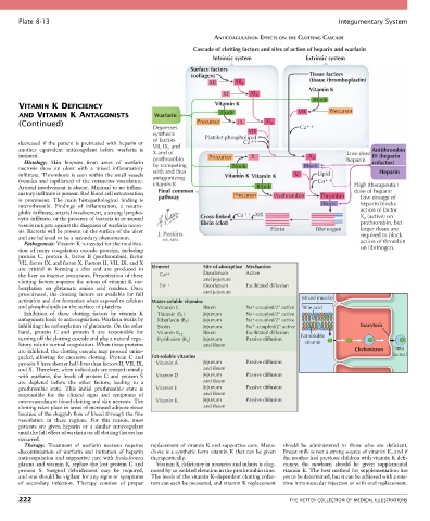
Plate 8-13                                                                                            Integumentary System

                                                                     ANTICOAGULATION EFFECTS ON THE CLOTTING CASCADE
                                                              Cascade of clotting factors and sites of action of heparin and warfarin
                                                                     Intrinsic system             Extrinsic system
                                                              Surface factors
                                                              (collagen)                           Tissue factors
                                                                    XII     XII a                  (tissue thromboplastin)
                                                                                                   Vitamin K
                                                                        XI       XI a
                                                                                                     Block
       VITAMIN K DEFICIENCY                                          Vitamin K                            Precursor
       AND VITAMIN K ANTAGONISTS                  Warfarin             Block                    VII
       (Continued)                                              Precursor    IX       XI a
                                                  Depresses                                     Ca
                                                  synthesis                     VIII
                                                  of factors      Platelet phospholipid
       decreased  if the  patient  is  pretreated  with heparin  or   VII, IX, and  Ca
       another  equivalent  anticoagulant  before  warfarin  is   X and of                                             Antithrombin
       initiated.                                 prothrombin      Precursor     X                 X a         Low-dose  III (heparin
         Histology:  Skin  biopsies  from  areas  of  warfarin                                                 heparin  cofactor)
       necrosis  show  an  ulcer  with  a  mixed  inflammatory     by competing  Block            Block
                                                  with and thus
       infiltrate. Thrombosis is seen within the small vessels   antagonizing  Vitamin K Vitamin K  V  Lipid              Heparin
       (venules and capillaries) of the cutaneous vasculature.   vitamin K                            Ca
       Arterial involvement is absent. Minimal to no inflam-                       Block                          High (therapeutic)
       matory infiltrate is present. Red blood cell extravasation   Final common   Precursor  Prothrombin  Thrombin  dose of heparin
       is  prominent.  The  main  histopathological  finding  is   pathway                                          Low dosage of
       microthrombi.  Findings  of  inflammation,  a  neutro-                                          Block        heparin blocks
       philic infiltrate, arterial involvement, a strong lympho-            Ca    XIII                              action of factor
       cytic infiltrate, or the presence of bacteria in or around   Cross-linked                                    X a (active) on
       vessels mitigate against the diagnosis of warfarin necro-  fibrin (clot)                                     prothrombin, but
       sis. Bacteria will be present on the surface of the ulcer                       Fibrin        Fibrinogen     larger doses are
       and are believed to be a secondary phenomenon.                                                               required to block
         Pathogenesis: Vitamin K is needed for the modifica-                                                        action of thrombin
       tion  of  many  coagulation  cascade  proteins,  including                                                   on fibrinogen.
       protein  C,  protein  S,  factor  II  (prothrombin),  factor
       VII, factor IX, and factor X. Factors II, VII, IX, and X
       are  critical  in  forming  a  clot  and  are  produced  in     Element  Site of absorption Mechanism
       the liver as inactive precursors. Preactivation of these         Ca ++  Duodenum  Active
       clotting  factors  requires  the  action  of  vitamin  K  car-  ++  and jejunum
       boxylation  on  glutamate  amino  acid  residues.  Once   Fe  Duodenum  Facilitated diffusion
       preactivated,  the  clotting  factors  are  available  for  full   and jejunum
       activation and clot formation when exposed to calcium   Water-soluble vitamins            Mixed micelle
       and phospholipids on the surface of platelets.      Vitamin C  Ileum    Na + -coupled/2° active  Bile acid
         Inhibition  of  these  clotting  factors  by  vitamin  K       Thiamin (B )  Jejunum  Na -coupled/2° active
                                                                                 +
                                                           1
       antagonists leads to anticoagulation. Warfarin works by       Riboflavin (B )  Jejunum  Na -coupled/2° active
                                                                                 +
                                                            2
       inhibiting the carboxylation of glutamate. On the other       Biotin  Jejunum  Na -coupled/2° active         Exocytosis
                                                                                 +
       hand,  protein  C  and  protein  S  are  responsible  for       Vitamin B 12  Ileum  Facilitated diffusion
       turning off the clotting cascade and play a natural regu-      Pyridoxine (B )  Jejunum  Passive diffusion  Fat-soluble
                                                            6
       latory role in normal coagulation. When these proteins     and ileum                       vitamin
       are inhibited, the clotting cascade may proceed unim-                                                      Chylomicron  Into
       peded,  allowing  for  excessive  clotting.  Protein  C  and   Fat-soluble vitamins                                    lacteal
       protein S have shorter half-lives than factors II, VII, IX,      Vitamin A  Jejunum  Passive diffusion
       and X. Therefore, when individuals are treated initially   and ileum
       with  warfarin,  the  levels  of  protein  C  and  protein  S        Vitamin D  Jejunum  Passive diffusion
       are  depleted  before  the  other  factors,  leading  to  a     and ileum
       prothrombic  state.  This  initial  prothrombic  state  is      Vitamin E  Jejunum  Passive diffusion
       responsible  for  the  clinical  signs  and  symptoms  of   and ileum
       microvasculature blood clotting and skin necrosis. The      Vitamin K  Jejunum  Passive diffusion
       clotting takes place in areas of increased adipose tissue   and ileum
       because of the sluggish flow of blood through the fine
       vasculature  in  these  regions.  For  this  reason,  most
       patients  are  given  heparin  or  a  similar  anticoagulant
       until the full effect of warfarin on all clotting factors has
       occurred.
         Therapy:  Treatment  of  warfarin  necrosis  requires   replacement of vitamin K and supportive care. Mena-  should  be  administered  to  those  who  are  deficient.
       discontinuation  of  warfarin  and  initiation  of  heparin   dione is a synthetic form vitamin K that can be given   Breast milk is not a strong source of vitamin K, and if
       anticoagulation and supportive care with fresh-frozen   therapeutically.            the mother had previous children with vitamin K defi-
       plasma and vitamin K replace the lost protein C and   Vitamin K deficiency in neonates and infants is diag-  ciency,  the  newborn  should  be  given  supplemental
       protein  S.  Surgical  debridement  may  be  required,     nosed by an isolated elevation in the prothrombin time.   vitamin K. The best method for supplementation has
       and one should be vigilant for any signs or symptoms   The levels of the vitamin K–dependent clotting cofac-  yet to be determined, but it can be achieved with a one-
       of  secondary  infection.  Therapy  consists  of  proper   tors can each be measured, and vitamin K replacement   time intramuscular injection or with oral replacement.

       222                                                                                   THE NETTER COLLECTION OF MEDICAL ILLUSTRATIONS