
Page 234 - The Netter Collection of Medical Illustrations - Integumentary System_ Volume 4 ( PDFDrive )
P. 234
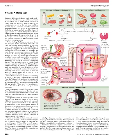
Plate 8-11                                                                                            Integumentary System

                                                        Principal food sources of vitamin A       Principal food sources of b-carotene
       VITAMIN A DEFICIENCY


       Vitamin A deficiency, also known as phrynoderma, is a   Butter   Egg yolk   Cod        Carrots       Tomatoes    Pimentos
       multisystem disorder caused by a deficiency of vitamin                     Liver
       A,  either  from  lack  of  intake  or  from  a  decrease  in               Oil
       normal absorption. Vitamin A is a fat-soluble essential                   Certain
       vitamin  that  is  stored  in  the  fatty  tissue  and  liver.   Milk  Liver  Kidneys  fish oils  Sweet  Apricots,  Leafy vegetables
       Humans require a nutritional source for this vitamin.                                    potatoes  yellow peaches
       Foods high in vitamin A include all yellow vegetables          CH 3   CH 3      CH            CH
       (including carrots), green leafy vegetables, liver, milk,          C              3             3
       eggs, tomatoes, and fish oils. Many other food staples         H 2 C  C  CH  CH  C  CH  CH  CH  C  CH 2  OH
       contain vitamin A. Hippocrates may have been the first         H 2 C  C  CH
       to describe vitamin A deficiency and a therapy for it.             C      3
       However, it was not until the early twentieth century   Vitamin A  H 2
       that scientists recognized the different forms of vitamin   Ester  Vitamin A                         β−carotene
       A and its carotene precursors.                                                                                    Thyroid
         Clinical Findings: Night blindness is one of the ear-                                                           promotes
       liest findings in vitamin A deficiency. Vitamin A is cru-                                                         conversion
       cially  important  for  proper  functioning  of  the  retinal   Bile aids  Thoracic             Bloodstream       of β−carotene
       rods, through production of rhodopsin. Rhodopsin is   absorption           duct                                   to vitamin A
       the primary rod pigment that makes visual adaption in   of β−carotene
       the dark possible. Xerophthalmia (dry eyes) often pre-                                            Esterase
       cedes the night blindness and is typically the first sign
       of  vitamin  A  deficiency,  although  this  sign  is  neither
       sensitive or specific. As the deficiency progresses, the                                               Liver     Mobil-
       xerophthalmia may result in corneal dryness, abrasions,           Pancreatic                   Storage           ization
       ulceration, and keratomalacia, which leads to blindness.          secretions
       Bitot’s spots can be seen on the lateral conjunctiva of           promote                                β−carotene
       the eye. These are highly specific for vitamin A defi-            hydrolysis                   Vitamin   converted
       ciency and appear as stuck-on foamy white papules and             of vitamin                   A ester-  to vitamin A
       plaques  that  cannot  be  removed  by  swabbing.  Bitot’s        A esters                     ified
       spots  are  caused  by  abnormal  keratinization  of  the
       conjunctival epithelium. It is estimated that vitamin A   Vitamin A
       deficiency  is  one  of  the  leading  causes  of  vision  loss   and β −carotene
       worldwide.  Growth  impairment  in  children  can  be   absorbed        Vitamin A
       caused by vitamin A deficiency.                                         esterified
         Phrynoderma is the name given to the skin findings
       in  vitamin  A  deficiency.  Phrynoderma  literally  means   Mineral oil
       “toad-like” skin, and it is manifested by hyperkeratotic   dissolves and
       follicle-based  papules.  The  skin  is  dry  and  rough.   carries off  Intestinal epithelial cell
       Patients with vitamin A deficiency may also have chei-  vitamin A
       litis  and  glossitis.  These  latter  two  conditions  are     in stool
       nonspecific  and  can  be  seen  in  a  variety  of  vitamin
       deficiencies.                                                       Principal deficiency manifestations
         Hypervitaminosis A can result from excessive vitamin
       A supplementation. It manifests as dry skin, hair loss,
       joint aches, bone pain, and headaches. Vitamin A can
       cause  birth  defects  when  taken  in  high  doses  during
       pregnancy.
         Pathogenesis:  Vitamin  A  deficiency  in  the  United
       States  is  most  frequently  caused  by  strange  dietary
       habits that avoid foods rich in vitamin A. Other condi-
       tions  may  predispose  individuals  to  this  deficiency,   Xerophthalmia (corneal
       including  cystic  fibrosis,  because  of  the  difficulty  in   abrasions and ulcerations)
       absorption of fat-soluble vitamins. Short gut syndrome   Bitot’s spots (stuck-on  Night blindness  Growth  Hyperkeratotic
       that  occurs  after  bariatric  surgery  may  also  lead  to   foamy white plaques)  (effect on retinal rods)  impairment follicular-based
       vitamin  A  deficiency.  Proper  production  of  bile  acids                                              papule (phrynoderma)
       and  pancreatic  enzymes  is  required  for  absorption  of
       vitamin A. Severe liver disease may result in functional
       vitamin  A  deficiency,  because  the  liver  is  required  to
       convert carotene into vitamin A.
         Vitamin A is found in foods predominantly as retinol   Histology:  Cutaneous  biopsies  are  nonspecific  but   vision has been shown to respond to therapy in some
       or β-carotene. Vitamin A is critical for nuclear signal-  may suggest a nutritional deficiency. There is pallor of   cases. Once blindness occurs, however, the only hope
       ing, through binding to its nuclear receptors, the reti-  the upper epidermis. Hyperkeratotic plugs are seen in   for vision is with corneal transplantation. Most cases in
       noic acid receptors (RARs) and the retinoid X receptors   follicles, with minimal to no inflammatory infiltrate.  North America and Europe are caused by poor absorp-
       (RXRs). Once this binding occurs, the resulting com-  Treatment:  Treatment  requires  replacement  of   tion  due  to  an  underlying  cause,  and  the  advice  of  a
       plexes can affect the transcription of various gene prod-  vitamin A and probably other essential vitamins in the   nutritionist who is an expert in malabsorption is indi-
       ucts.  The  vitamin  is  responsible  for  maturation  and   patient’s diet. The eye changes may be permanent, but   cated. These patients may require long-term replace-
       proliferation of epithelial cells.        the cutaneous findings respond well. Loss of only night   ment and monitoring of their vitamin A levels.

       220                                                                                   THE NETTER COLLECTION OF MEDICAL ILLUSTRATIONS