
Page 237 - The Netter Collection of Medical Illustrations - Integumentary System_ Volume 4 ( PDFDrive )
P. 237
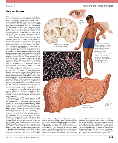
Plate 8-14                                                                                Nutritional and Metabolic Diseases


        WILSON’S DISEASE


        Wilson’s disease, also known as hepatolenticular degen-
        eration, is a disorder caused by a defect in copper metab-
        olism. The disease is rare, with a worldwide incidence                              Kayser-
        of approximately 1 in 18,000. It is an autosomal reces-                             Fleischer
        sive condition that is caused by a defect in the ATP7B                              ring
        gene, which is located on the long arm of chromosome
        13.  The  product  of  this  gene  is  responsible  for  the
        proper transport of copper. The main clinical findings
        relate to nervous system involvement and liver disease.
        Wilson’s disease has a variable phenotype depending on
        the  specific  genetic  mutation.  Cutaneous  disease  and
        ophthalmological disease are frequently seen.
          Clinical  Findings:  Wilson’s  disease  equally  affects
        males and females, and its incidence varies among pop-
        ulations. The usual age at onset is in the first 2 decades
        of life. Liver disease and central nervous system (CNS)
        disease are often the first signs. Patients may present   Degenerative changes                         Adolescents more likely
                                                                                                               have generalized dystonia
        with  unexplained  hepatomegaly,  cirrhosis,  and  end-  in lenticular nuclei                          with neck (torticollis) and
        stage liver disease. The CNS findings can manifest in                                                  face (grimacing) effects,
        various patterns. Mild to severe psychiatric symptoms                                                  occasionally focal;
        of depression and mood lability are common; the mani-                                                  hypertonicity and
        festations in some patients may approach the diagnosis                                                 choreoathetosis may coexist
        of schizophrenia. Impaired cognition and memory are
        frequently seen and may lead to early dementia. Extra-                                                 Adults more likely have
                                                                                                               coarse, proximal “wing
        pyramidal  features  are  always  present  and  include                                                beating” or “chest beating”
        tremor and rigidity. The tremor has been described as                                                  tremor, masked facies,
        a “wing-beating” tremor of the shoulder girdle. Brady-                                                 and dysarthric speech
        kinesia  is  invariably  a  part  of  the  disease.  Ataxia  and
        chorea, along with dysfunction of normal motor coor-
        dination, are evident as time progresses.
          The cutaneous findings, when present in conjunction
        with  liver  and  CNS  disease,  can  make  the  diagnosis.
        Patients  have  varying  amounts  of  pretibial  hyperpig-
        mentation,  the  cause  of  which  is  poorly  understood.
        Rarely, patients present with a blue discoloration to the
        lunula of the nail. The most pathognomonic sign is the
        presence  of  Kayser-Fleischer  rings  on  the  cornea.  A
        Kayser-Fleischer ring is a yellow to orange-brown ring
        around the iris. It represents an abnormal accumulation
        of  copper  in  Descemet’s  membrane  of  the  cornea.  A
        slit-lamp  examination  is  required  to  appreciate  this
        clinical finding, which is unique to Wilson’s disease.
          Laboratory testing is required to confirm the diag-
        nosis.  A  hallmark  is  a  decreased  ceruloplasmin  level.
        The actual ceruloplasmin protein is not defective in any
        manner. Urinary copper excretion is elevated to more
        than 100 µg/day.
          Pathogenesis: The ATP7B gene is mutated in Wil-
        son’s disease, and this leads to the systemic and cutane-
        ous  manifestations  of  the  disease.  The  ATP7B  gene
        encodes the P-type adenosine triphosphatase (ATPase)
        that  serves  as  a  metal-binding  and  metal-carrying                                  Postnecrotic
        protein.  This  P-type  ATPase  is  primarily  responsible                               type of cirrhosis
        for the transport of copper. When it is defective, copper
        builds up to abnormal levels in the liver, in the CNS,
        and  to  a  lesser  extent,  in  the  cornea.  Many  different
        mutations have been discovered in the ATP7B gene and
        are  responsible  for  the  different  phenotypes  seen.
        Homozygous  patients  and  compound  heterozygotes
        have completely different phenotypes with some over-  cirrhosis. Hydropic degeneration of individual hepato-  normal. CNS symptoms, if present at the time of trans-
        lapping  features.  Certain  mutations  lead  to  liver  and   cytes  is  seen  to  a  varying  degree,  depending  on  the   plantation, typically persist with minimal improvement
        CNS disease or to a predominance of one over the other.   timing of the biopsy. Special staining methods can high-  over time. While awaiting liver transplantation, patients
        The large number of mutations and the large size of the   light the elevated copper within the hepatocytes.  are usually treated with a combination of a low-copper
        gene make it difficult to analyze. The prevalence of the   Treatment:  The  only  cure  for  the  disease  is  liver   diet,  oral  zinc  supplementation,  and  D-penicillamine.
        different genetic mutations varies among populations.  transplantation.  This  procedure  is  becoming  more   Zinc competes with copper for absorption and decreases
          Histology: Skin biopsies are not helpful in the diag-  common and has led to excellent therapeutic responses.   the amount of copper absorbed from the gastrointesti-
        nosis.  Biopsies  of  the  liver  show  varying  degrees     The transplanted normal liver produces adequate levels   nal  tract.  D-Penicillamine  is  a  copper-chelating  agent
        of  portal  inflammation  and  fibrosis  with  eventual     of  the  P-type  ATPase  to  bring  the  copper  levels  to   that helps to lower serum and tissue copper levels.


        THE NETTER COLLECTION OF MEDICAL ILLUSTRATIONS                                                                          223