
Page 241 - The Netter Collection of Medical Illustrations - Integumentary System_ Volume 4 ( PDFDrive )
P. 241
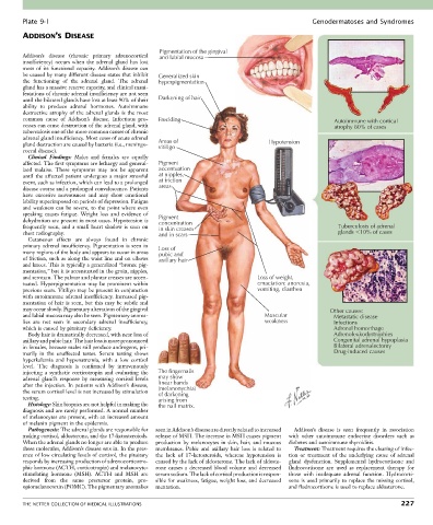
Plate 9-1                                                                                   Genodermatoses and Syndromes
        ADDISON’S DISEASE


                                                   Pigmentation of the gingival
        Addison’s  disease  (chronic  primary  adrenocortical   and labial mucosa
        insufficiency)  occurs  when  the  adrenal  gland  has  lost
        most  of  its  functional  capacity.  Addison’s  disease  can
        be caused by many different disease states that inhibit   Generalized skin
        the  functioning  of  the  adrenal  gland.  The  adrenal     hyperpigmentation
        gland has a massive reserve capacity, and clinical mani-
        festations of chronic adrenal insufficiency are not seen
        until the bilateral glands have lost at least 90% of their   Darkening of hair
        ability  to  produce  adrenal  hormones.  Autoimmune
        destructive  atrophy  of  the  adrenal  glands  is  the  most
        common  cause  of  Addison’s  disease.  Infectious  pro-  Freckling                                Autoimmune with cortical
        cesses can cause destruction of the adrenal gland, with                                            atrophy 80% of cases
        tuberculosis one of the more common causes of chronic
        adrenal gland insufficiency. Most cases of acute adrenal   Areas of
        gland destruction are caused by bacteria (i.e., meningo-  vitiligo            Hypotension
        coccal disease).
          Clinical  Findings:  Males  and  females  are  equally
        affected. The first symptoms are lethargy and general-  Pigment
        ized  malaise.  These  symptoms  may  not  be  apparent   accentuation
        until  the  affected  patient  undergoes  a  major  stressful   at nipples,
        event, such as infection, which can lead to a prolonged   at friction
        disease course and a prolonged convalescence. Patients   areas
        have  excessive  nervousness  and  may  show  emotional
        lability superimposed on periods of depression. Fatigue
        and weakness can be severe, to the point where even
        speaking  causes  fatigue.  Weight  loss  and  evidence  of   Pigment
        dehydration are present in most cases. Hypotension is   concentration
        frequently  seen,  and  a  small  heart  shadow  is  seen  on   in skin creases                     Tuberculosis of adrenal
        chest radiography.                         and in scars                                             glands  10% of cases
          Cutaneous  effects  are  always  found  in  chronic
        primary adrenal insufficiency. Pigmentation is seen in   Loss of
        many regions of the body and appears to occur in areas   pubic and
        of friction, such as along the waist line and on elbows   axillary hair
        and knees. This is typically a generalized “bronze pig-
        mentation,” but it is accentuated in the groin, nipples,
        and scrotum. The palmar and plantar creases are accen-                    Loss of weight,
        tuated. Hyperpigmentation may be prominent within                         emaciation: anorexia,
        previous scars. Vitiligo may be present in conjunction                    vomiting, diarrhea
        with autoimmune adrenal insufficiency. Increased pig-
        mentation of hair is seen, but this may be subtle and
        may occur slowly. Pigmentary alterations of the gingival                                         Other causes:
        and labial mucosa may also be seen. Pigmentary anoma-                        Muscular             Metastatic disease
        lies  are  not  seen  in  secondary  adrenal  insufficiency,                 weakness             Infections
        which is caused by pituitary deficiency.                                                          Adrenal hemorrhage
          Body hair is dramatically decreased, with near loss of                                          Adrenoleukodystrophies
        axillary and pubic hair. The hair loss is more pronounced                                         Congenital adrenal hypoplasia
        in females, because males still produce androgens, pri-                                           Bilateral adrenalectomy
        marily  in  the  unaffected  testes.  Serum  testing  shows                                       Drug-induced causes
        hyperkalemia  and  hyponatremia,  with  a  low  cortisol
        level.  The  diagnosis  is  confirmed  by  intravenously
        injecting  a  synthetic  corticotropin  and  evaluating  the   The fingernails
        adrenal  gland’s  response  by  measuring  cortisol  levels   may show
        after the injection. In patients with Addison’s disease,     linear bands
                                                   (melanonychia)
        the serum cortisol level is not increased by stimulation   of darkening
        testing.                                   arising from
          Histology: Skin biopsies are not helpful in making the   the nail matrix.
        diagnosis and are rarely performed. A normal number
        of melanocytes are present, with an increased amount
        of melanin pigment in the epidermis.
          Pathogenesis: The adrenal glands are responsible for   seen in Addison’s disease are directly related to increased   Addison’s  disease  is  seen  frequently  in  association
        making cortisol, aldosterone, and the 17-ketosteroiods.   release of MSH. The increase in MSH causes pigment   with  other  autoimmune  endocrine  disorders  such  as
        When the adrenal glands no longer are able to produce   production  by  melanocytes  in  skin,  hair,  and  mucous   diabetes and autoimmune thyroiditis.
        these molecules, Addison’s disease sets in. In the pres-  membranes.  Pubic  and  axillary  hair  loss  is  related  to     Treatment: Treatment requires the clearing of infec-
        ence of low circulating levels of cortisol, the pituitary   the  lack  of  17-ketosteroids,  whereas  hypotension  is   tion  or  treatment  of  the  underlying  cause  of  adrenal
        responds by increasing production of adrenocorticotro-  caused by the lack of aldosterone. The lack of aldoste-  gland  dysfunction.  Supplemental  hydrocortisone  and
        phic hormone (ACTH, corticotropin) and melanocyte-  rone  causes  a  decreased  blood  volume  and  decreased   fludrocortisone  are  used  as  replacement  therapy  for
        stimulating  hormone  (MSH).  ACTH  and  MSH  are   serum sodium. The lack of cortisol production is respon-  those  with  inadequate  adrenal  function.  Hydrocorti-
        derived  from  the  same  precursor  protein,  pro-  sible for weakness, fatigue, weight loss, and decreased   sone is used primarily to replace the missing cortisol,
        opiomelanocortin (POMC). The pigmentary anomalies   mentation.                      and fludrocortisone is used to replace aldosterone.


        THE NETTER COLLECTION OF MEDICAL ILLUSTRATIONS                                                                          227