
Page 242 - The Netter Collection of Medical Illustrations - Integumentary System_ Volume 4 ( PDFDrive )
P. 242
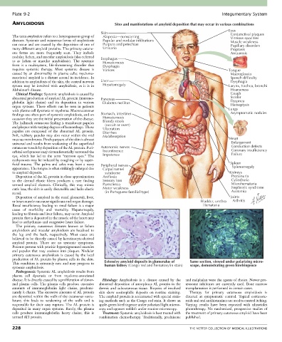
Plate 9-2                                                                                             Integumentary System
       AMYLOIDOSIS                                       Sites and manifestations of amyloid deposition that may occur in various combinations
                                                                                                                Eyes
                                                  Skin
       The term amyloidosis refers to a heterogeneous group of   Alopecia—nonscarring                            Conjunctival plaques
                                                                                                                 Vitreous opacities
       diseases. Systemic and cutaneous forms of amyloidosis   Papular and nodular infiltrations                 Muscle weakness
       can occur and are caused by the deposition of one of   Purpura and petechiae                              Pupillary disorders
       many different amyloid proteins. The primary cutane-  Urticaria                                           Proptosis
       ous  forms  are  more  frequently  seen.  They  include                                                   Amaurosis
       nodular, lichen, and macular amyloidosis (also referred   Esophagus
       to  as  lichen  or  macular  amyloidosis).  The  systemic     Hematemesis
       form  is  a  multisystem,  life-threatening  disorder  that   Dysphagia
       requires  systemic  therapy.  Most  systemic  disease  is   Varices                                      Tongue
       caused  by  an  abnormality  in  plasma  cells;  myeloma-                                                 Macroglossia
       associated amyloid is a distant second in incidence. In                                                   Speech difficulty
       addition to amyloidosis of the skin, the central nervous   Liver                                          Dysphagia
       system  may  be  involved  with  amyloidosis,  as  it  is  in   Hepatomegaly                             Larynx, trachea, bronchi
       Alzheimer’s disease.                                                                                      Hoarseness
         Clinical Findings: Systemic amyloidosis is caused by                                                    Cough
       abnormal production of amyloid AL protein (immuno-  Pancreas                                              Stridor
       globulin  light  chains)  and  its  deposition  in  various   Diabetes mellitus                           Dyspnea
       organ  systems.  These  effects  can  be  seen  in  patients                                              Hemoptysis
       with plasma cell dyscrasia or myeloma. Mucocutaneous                                                     Lungs
       findings are often part of systemic amyloidosis, and on   Stomach, intestines                             Asymptomatic nodules
       occasion they are the initial presentation of the disease.   Hematemesis
       The hallmark cutaneous finding is translucent papules   Bloody stools
                                                    (occult or overt)
       and plaques with varying degrees of hemorrhage. These   Ulceration
       papules  are  composed  of  the  abnormal  AL  protein.   Diarrhea
       Soft,  rubbery  papules  may  also  occur  within  the  oral   Malabsorption
       mucous membranes. Pinch purpura of the skin is almost                                                    Heart
       universal and results from weakening of the superficial                                                   Enlargement
       cutaneous vessels by deposition of the AL protein. Peri-  Autonomic nerves                                Conduction defects
       orbital ecchymoses may circumferentially surround the   Incontinence                                      Coronary insufficiency
       eye,  which  has  led  to  the  term  “raccoon  eyes.”  The   Impotence                                   Failure
       ecchymoses may be induced by coughing or by super-
       ficial  trauma.  The  palms  and  soles  may  have  a  waxy   Peripheral nerves                           Spleen
       appearance. The tongue is often strikingly enlarged due   Carpal tunnel                                    Splenomegaly
       to amyloid deposits.                         syndrome                                                    Kidneys
         Deposition of the AL protein in close approximation   Areflexia                                         Proteinuria
       to  the  dermal  elastic  fibers  produces  a  rare  finding   Sensory loss                               Cylindruria
       termed  amyloid  elastosis.  Clinically,  this  may  mimic   Paresthesia                                  Microhematuria
       cutis laxa; the skin is easily distensible and lacks elastic   Motor weakness                             Nephrotic syndrome
       recoil.                                      (in Portuguese familial type)                                Azotemia
         Deposition of amyloid in the renal glomeruli, liver,                                                    Joints
       or heart muscle can cause significant end-organ damage.                                    Bladder, urethra  Arthritis
       Renal insufficiency leading to renal failure is a major                                     Hematuria
       cause  of  morbidity  and  mortality.  Hepatomegaly,
       leading to fibrosis and liver failure, may occur. Amyloid
       protein that is deposited in the muscle of the heart may
       lead to arrhythmias and congestive heart failure.
         The  primary  cutaneous  diseases  known  as  lichen
       amyloidosis  and  macular  amyloidosis  are  localized  to
       the  leg  and  the  back,  respectively.  Most  cases  are
       believed to be directly caused by keratinocyte-derived
       amyloid  protein.  There  are  no  systemic  symptoms.
       Patients present with pruritic hyperpigmented macules
       and  papules  that  may  coalesce  into  plaques.  Nodular
       primary  cutaneous  amyloidosis  is  caused  by  the  local
       production of AL protein by plasma cells in the skin.
       This condition is extremely rare and may progress to   Extensive amyloid deposits in glomerulus of  Same section, viewed under polarizing micro-
                                                                                             scope, demonstrating green birefringence
                                                    human kidney (Congo red and hematoxylin stain)
       systemic amyloidosis.
         Pathogenesis: Systemic AL amyloidosis results from
       plasma  cell  dyscrasia  or  from  myeloma-associated
       disease. It is directly caused by a proliferation of abnor-  Histology:  Amyloidosis  is  a  disease  caused  by  the   and melphalan were the agents of choice. Newer pro-
       mal  plasma  cells.  The  plasma  cells  produce  excessive   abnormal deposition of amorphous AL protein in the   teosome  inhibitors  are  currently  used.  Bone  marrow
       amounts  of  immunoglobulin  light  chains,  predomi-  dermis  and  subcutaneous  tissue.  Biopsies  of  involved   transplantation is performed in certain cases.
       nantly λ chains. The excessive amounts of AL protein   skin  show  eosinophilic  deposits  on  routine  staining.   Therapy  for  primary  cutaneous  amyloidosis  is
       are deposited within the walls of the cutaneous vascu-  The amyloid protein is accentuated with special stain-  directed  at  symptomatic  control.  Topical  corticoste-
       lature;  this  leads  to  weakening  of  the  walls  and  is   ing methods such as the Congo red stain. It shows an   roids and oral antihistamines are used to control itching.
       responsible for their easy rupture. The AL protein is   apple-green birefringence under polarized light micros-  Varying  results  have  been  reported  with  ultraviolet
       deposited  in  many  organ  systems.  Rarely,  the  plasma   copy and appears reddish under routine microscopy.  phototherapy.  No  randomized,  prospective  studies  of
       cells  produce  immunoglobulin  heavy  chains;  this  is   Treatment: Systemic amyloidosis is best treated with   the treatment of primary cutaneous amyloid have been
       termed AH protein.                        combination  chemotherapy.  Traditionally,  prednisone   published.

       228                                                                                   THE NETTER COLLECTION OF MEDICAL ILLUSTRATIONS