
Page 54 - The Netter Collection of Medical Illustrations - Integumentary System_ Volume 4 ( PDFDrive )
P. 54
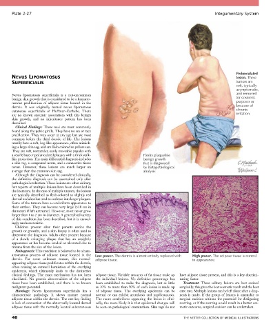
Plate 2-27                                                                                            Integumentary System


                                                                                                                       Pedunculated
       NEVUS LIPOMATOSUS                                                                                               lesion. These
       SUPERFICIALIS                                                                                                   tumors are
                                                                                                                       soft, typically
                                                                                                                       asymptomatic,
       Nevus  lipomatosus  superficialis  is  a  not-uncommon                                                          and removed
       benign skin growth that is considered to be a hamarto-                                                          for cosmetic
       matous  proliferation  of  adipose  tissue  located  in  the                                                    purposes or
       dermis.  It  was  originally  named  nevus  lipomatosus                                                         because of
       cutaneous  superficialis  of  Hoffman-Zurhelle.  There                                                          chronic
       are  no  known  systemic  associations  with  this  benign                                                      irritation.
       skin  growth,  and  no  inheritance  pattern  has  been
       described.
         Clinical  Findings:  These  nevi  are  most  commonly
       found along the pelvic girdle. They have no sex or race
       predilection. They may occur at any age but are most
       common  before  the  third  decade  of  life.  The  lesions
       usually have a soft, bag-like appearance, often mimick-
       ing a large skin tag, and are flesh colored to yellow-tan.
       They are soft, nontender, easily moveable papules with
       a sessile base or pedunculated plaques with a thick stalk-        Fleshy plaquelike
       like projection. The main differential diagnosis includes         benign growth
       a skin tag, a compound nevus, and a connective tissue             that is diagnosed
       nevus.  However,  these  lesions  are  much  larger  on           by histopathological
       average than the common skin tag.                                 analysis
         Although the diagnosis can be considered clinically,
       the  definitive  diagnosis  can  be  ascertained  only  after
       pathological evaluation. These lesions are often solitary,
       but reports of multiple lesions have been described in
       the literature. In the case of multiple tumors, the lesions
       are typically described as flesh-colored to slightly red
       dermal nodules that tend to coalesce into larger plaques.
       Some of the tumors have a cerebriform appearance to
       their surface. They can become very large (>10 cm in
       diameter) if left untreated. However, most never grow
       larger than 1 to 2 cm in diameter. A generalized variety
       of this condition has been described, but it is exceed-
       ingly uncharacteristic.
         Children  present  after  their  parents  notice  the
       growth or growths, and a skin biopsy is often used to
       determine the diagnosis. Adults often present because
       of  a  slowly  enlarging  plaque  that  has  an  unsightly
       appearance or has become eroded or ulcerated due to
       trauma from the size of the lesion.
         Pathogenesis: This condition is believed to be a ham-
       artomatous  process  of  adipose  tissue  located  in  the   Low power. The dermis is almost entirely replaced with  High power. The adipose tissue is normal
       dermis.  For  some  unknown  reason,  this  normal-  adipose tissue.                      in appearance.
       appearing adipose tissue proliferates within the dermis,
       often  causing  an  outward  herniation  of  the  overlying
       epidermis,  which  ultimately  leads  to  the  distinctive
       clinical  findings.  The  exact  mechanism  has  not  been   adipose tissue. Variable amounts of fat tissue make up   have adipose tissue present, and this is a key discrimi-
       elucidated.  No  genetic  abnormalities  of  the  adipose   the  individual  lesions.  No  definitive  percentage  has   nating  factor.
       tissue  have  been  established,  and  there  is  no  known   been  established  to  make  the  diagnosis,  but  as  little   Treatment:  These  solitary  lesions  are  best  excised
       malignant potential.                      as 10% to more than 50% of each lesion is made up   surgically; this gives the best cosmetic result and the best
         Histology:  Nevus  lipomatosus  superficialis  has  a   of  adipose  tissue.  The  overlying  epidermis  can  be   cure rate. Multiple lesions can be left alone after a diag-
       characteristic  pathology.  It  shows  mature  normal   normal  or  can  exhibit  acanthosis  and  papillomatosis.   nosis  is  made.  If  the  group  of  lesions  is  amenable  to
       adipose tissue within the dermis. The one key finding   The  more  cerebriform  appearing  the  lesion  is  clini-  surgical  excision  without  the  potential  for  disfiguring
       is lack of connection of the abnormally located dermal   cally, the more likely it is that epidermal changes will   scarring, or if the scarring would result in a better cos-
       adipose tissue with the normally located subcutaneous   be seen on pathological examination. Skin tags do not   metic outcome, surgical excision can be undertaken.

       40                                                                                    THE NETTER COLLECTION OF MEDICAL ILLUSTRATIONS