
Page 71 - The Netter Collection of Medical Illustrations - Integumentary System_ Volume 4 ( PDFDrive )
P. 71
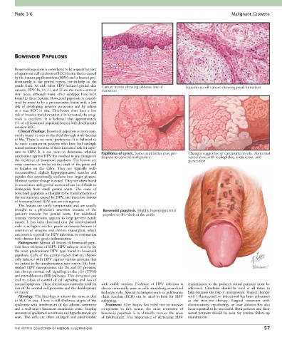
Plate 3-6                                                                                                Malignant Growths


        BOWENOID PAPULOSIS


        Bowenoid papulosis is considered to be a special variant
        of squamous cell carcinoma (SCC) in situ that is caused
        by the human papillomavirus (HPV) and is located pre-
        dominantly  in  the  genital  region,  particularly  on  the
        penile shaft. As with other HPV-induced genital skin   Cancer in situ showing oblique line of  Squamous cell cancer showing pearl formation
        cancers, HPV 16, 18, 31, and 33 are the more common   transition
        viral  types,  although  many  other  subtypes  have  been
        found in these lesions. Bowenoid papulosis is consid-
        ered by some to be a precancerous lesion with a low
        risk  of  developing  invasive  properties  and  by  others
        as  a  true  SCC  in  situ.  This  lesion  does  have  a  low
        risk of invasive transformation; if it is treated, the prog-
        nosis  is  excellent.  It  is  believed  that  approximately
        1% of all bowenoid papulosis lesions will develop into
        invasive SCC.
          Clinical Findings: Bowenoid papulosis is most com-
        monly found in men in the third through sixth decades
        of life. There is no racial preference. It is believed to
        be  more  common  in  patients  who  have  had  multiple
        sexual partners because of their increased risk for expo-
        sure  to  HPV.  It  is  too  soon  to  determine  whether     Papilloma of cervix. Some papillomas may pre-  Changes suggestive of carcinoma in situ. Abnormal
        vaccination against HPV has resulted in any changes in   dispose to cervical malignancy.  vasculature with leukoplakia, mosaicism, and
        the incidence of bowenoid papulosis. The lesions are                                punctation
        most common in males on the shaft of the penis and
        in  females  on  the  vulva.  They  are  typically  well-
        circumscribed,  slightly  hyperpigmented  macules  and
        papules that occasionally coalesce into larger plaques.
        Minimal surface change is noted. They are often found
        in association with genital warts and can be difficult to
        distinguish  from  small  genital  warts.  The  cause  of
        bowenoid papulosis is thought to be transformation of
        the keratinocyte caused by HPV, and therefore lesions
        of bowenoid shed HPV and are contagious.
          The lesions are rarely symptomatic and are usually
        brought  to  a  physician’s  attention  because  of  the   Bowenoid papulosis. Slightly hyperpigmented
        patient’s  concern  for  genital  warts.  For  undefined   papules on the shaft of the penis
        reasons,  circumcision  appears  to  help  prevent  penile
        cancer.  It  has  been  theorized  that  the  uncircumcised
        male is at higher risk for penile carcinoma because of
        retention  of  smegma  and  chronic  maceration,  which
        can provide a portal for HPV infection, in conjunction
        with chronic low-grade inflammation.
          Pathogenesis: Almost all lesions of bowenoid papu-
        losis have evidence of HPV. HPV subtype 16 is by far
        the  most  predominant  HPV  type  found  in  bowenoid
        papulosis. Cells of the genital region that are chroni-
        cally infected with HPV express various proteins that
        are critical in the transformation into cancer. The best-
        studied  HPV  oncoproteins,  the  E6  and  E7  proteins,
        can  disrupt  normal  cell  signaling  in  the  p16  (TP16)
        and retinoblastoma (RB) pathways. This disruption can
        lead  to  a  loss  of  control  of  cell  signaling  and  loss  of
        normal apoptosis. These alterations eventually result in   with  visible  mitoses.  Evidence  of  HPV  infection  is   transmission  to  the  patient’s  sexual  partners  must  be
        loss of the normal cell processes and the development   almost  universally  seen  as  cells  mimicking  vacuolated   addressed.  Condoms  should  be  used  at  all  times  to
        of cancer.                                koilocyte cells. Special techniques such as polymerase   help decrease the risk of transmission. Topical therapy
          Histology: The histology is almost the same as that   chain  reaction  (PCR)  can  be  used  to  look  for  HPV   with 5-fluorouracil or imiquimod has been advocated
        of  SCC  in  situ.  There  is  full-thickness  atypia  of  the   subtyping.         as  the  first-line  therapy.  Surgical  treatment  with
        epidermis  with  involvement  of  the  adnexal  structures   Treatment:  After  biopsy  has  ruled  out  an  invasive   electrocautery,  cryotherapy,  or  laser  ablation  has  also
        and  a  well-intact  basement  membrane  zone.  Varying   component  to  this  tumor,  the  main  treatment  of   been reported to be successful. Both patients and their
        amounts of epidermal acanthosis and hyperkeratosis are   bowenoid  papulosis  is  to  clinically  remove  the  areas     sexual  partners  should  be  seen  for  routine  follow-up
        seen.  The  cells  are  often  enlarged  and  pleomorphic   of  involvement.  The  importance  of  decreasing  HPV   examinations.


        THE NETTER COLLECTION OF MEDICAL ILLUSTRATIONS                                                                           57