
Page 70 - The Netter Collection of Medical Illustrations - Integumentary System_ Volume 4 ( PDFDrive )
P. 70
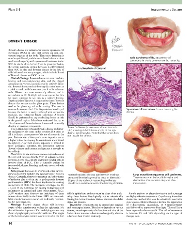
Plate 3-5                                                                                             Integumentary System


       BOWEN’S DISEASE


       Bowen’s disease is a variant of cutaneous squamous cell
       carcinoma  (SCC)  in  situ  that  occurs  on  non–sun-
       exposed  regions  of  the  body.  That  strict  definition  is
       not always followed, and the term Bowen’s disease is often                             Early carcinoma of lip. Squamous cell
       used interchangeably with squamous cell carcinoma in situ.                             carcinoma in situ is common on the lower lip.
       SCC in situ is often derived from its precursor lesion,
       the actinic keratosis. Actinic keratosis is differentiated   Erythroplasia of Queyrat
       from  SCC  in  situ  and  Bowen’s  disease  by  its  lack  of
       full-thickness keratinocyte atypia, which is the hallmark
       of Bowen’s disease and SCC in situ.
         Clinical Findings: Bowen’s disease can occur on hair-
       bearing  and  non–hair-bearing  skin,  and  the  clinical
       appearance in various locations can be entirely differ-
       ent. Bowen’s disease on hair-bearing skin often starts as
       a  pink  to  red,  well-demarcated  patch  with  adherent
       scale.  Women  are  most  commonly  affected,  and  it
       occurs later in life. Multiple lesions can occur, but it is
       far  more  common  to  see  this  as  a  solitary  finding.
       Erythroplasia of Queyrat is a regional variant of Bowen’s
       disease  that  occurs  on  the  glans  penis.  These  lesions
       tend  to  be  glistening  red  with  crusting.  The  area  is
       often well circumscribed. The diagnosis is often delayed                            Squamous cell carcinoma. Tumor invading the
       because  the  lesion  is  easily  confused  with  dermatitis,                       dermis.
       psoriasis,  and  cutaneous  fungal  infections.  A  biopsy
       should be performed on any nonhealing lesion or rash
       in the genital region. It has been estimated that up to
       5% of untreated Bowen’s disease lesions will eventually
       develop an invasive component.            Bowen’s disease (squamous cell carcinoma in
         The relationship between Bowen’s disease and inter-
       nal malignancy has come under scrutiny; if it exists at   situ) showing full-thickness atypia of the epi-
                                                 dermal keratinocytes. Note that the tumor does
       all, it is likely a consequence of the use of arsenic in the   not invade the dermis.
       past. Patients with a history of arsenic ingestion are at
       a higher risk of developing Bowen’s disease and internal
       malignancy.  Now  that  arsenic  exposure  is  limited  in
       most  developed  countries,  the  association  between
       Bowen’s disease and internal malignancy is thought to
       be unlikely.
         Most SCCs in situ are found on sun-exposed areas of
       the skin and develop directly from an adjacent actinic
       keratosis. Some SCCs in situ eventually develop into an
       invasive  form  of  SCC.  This  is  clinically  evident  by
       increased thickness, bleeding, and pain associated with
       the lesion.
         Pathogenesis: Exposure to arsenic and other carcino-
       gens has been implicated in the development of Bowen’s   Perianal Bowen’s disease can have an insidious  Large crateriform squamous cell carcinoma.
       disease. Certainly, ultraviolet radiation and other forms   onset and be misdiagnosed as tinea or dermatitis.  These tumors can be locally invasive and
       of radiation play a role in the its pathogenesis. Human   Biopsy of any rash not responding to therapy  destructive. On occasion they can also
       papillomavirus (HPV) has been implicated in causing   should be a consideration for the treating clinician.   metastasize.
       many forms of SCC. The oncogenic viral types 16, 18,
       31, and 33 are notorious for causing mutagenesis and
       malignancy in cervical and some other genital SCCs.
       HPV  vaccines  may  decrease  the  incidence  of  these   follicle epithelium, and care must be taken when evalu-  Simple  excision  or  electrodessication  and  curettage
       tumors dramatically in the future. HPV can cause cel-  ating  these  lesions  histologically  not  to  mistake  this   are highly effective treatments. Cryotherapy is another
       lular transformations to occur and is directly responsi-  finding for dermal invasion. Various amounts of cellular   destructive  method  that  can  be  selectively  used  with
       ble for tumorigenesis.                    atypia are present.                       good success. Medical therapies include the application
         Histology:  Bowen’s  disease  shows  full-thickness   Treatment: Treatments can be divided into surgical   of  5-fluorouracil,  imiquimod,  or  5-aminolevulinic
       atypia  of  the  keratinocytes  within  the  epidermis.  No   and nonsurgical forms. The choice depends on various   acid followed by exposure to blue light. These all have
       dermal invasion is present. The underlying dermis may   factors, most importantly the location and size of the   been reported to be successful. The risk of recurrence
       show a lymphocytic perivascular infiltrate. The atypia   lesion. Some tumors are best treated surgically, whereas   is  between  3%  and  10%  depending  on  the  type  of
       of the keratinocytes extends down to involve the hair   others are best treated medically.  therapy used.

       56                                                                                    THE NETTER COLLECTION OF MEDICAL ILLUSTRATIONS