
Page 69 - The Netter Collection of Medical Illustrations - Integumentary System_ Volume 4 ( PDFDrive )
P. 69
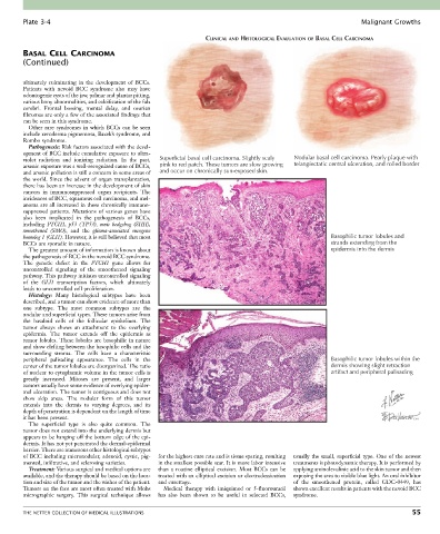
Plate 3-4                                                                                                Malignant Growths

                                                                 CLINICAL AND HISTOLOGICAL EVALUATION OF BASAL CELL CARCINOMA

        BASAL CELL CARCINOMA
        (Continued)


        ultimately  culminating  in  the  development  of  BCCs.
        Patients  with  nevoid  BCC  syndrome  also  may  have
        odontogenic cysts of the jaw, palmar and plantar pitting,
        various bony abnormalities, and calcification of the falx
        cerebri.  Frontal  bossing,  mental  delay,  and  ovarian
        fibromas are only a few of the associated findings that
        can be seen in this syndrome.
          Other  rare  syndromes  in  which  BCCs  can  be  seen
        include xeroderma pigmentosa, Bazek’s syndrome, and
        Rombo syndrome.
          Pathogenesis: Risk factors associated with the devel-
        opment of BCC include cumulative exposure to ultra-
        violet  radiation  and  ionizing  radiation.  In  the  past,   Superficial basal cell carcinoma. Slightly scaly  Nodular basal cell carcinoma. Pearly plaque with
        arsenic exposure was a well-recognized cause of BCCs,   pink to red patch. These tumors are slow growing  telangiectatic central ulceration, and rolled border
        and arsenic pollution is still a concern in some areas of   and occur on chronically sun-exposed skin.
        the world. Since the advent of organ transplantation,
        there has been an increase in the development of skin
        cancers  in  immunosuppressed  organ  recipients.  The
        incidences of BCC, squamous cell carcinoma, and mel-
        anoma are all increased in these chronically immuno-
        suppressed  patients.  Mutations  of  various  genes  have
        also  been  implicated  in  the  pathogenesis  of  BCCs,
        including  PTCH1,  p53  ( TP53),  sonic  hedgehog  (SHH),
        smoothened  (SMO),  and  the  glioma-associated  oncogene
        homolog 1 (GLI1). However, it is still believed that most                                       Basophilic tumor lobules and
        BCCs are sporadic in nature.                                                                    strands extending from the
          The greatest amount of information is known about                                             epidermis into the dermis
        the pathogenesis of BCC in the nevoid BCC syndrome.
        The  genetic  defect  in  the  PTCH1  gene  allows  for
        uncontrolled  signaling  of  the  smoothened  signaling
        pathway. This pathway initiates uncontrolled signaling
        of  the  GLI1  transcription  factors,  which  ultimately
        leads to uncontrolled cell proliferation.
          Histology:  Many  histological  subtypes  have  been
        described, and a tumor can show evidence of more than
        one  subtype.  The  most  common  subtypes  are  the
        nodular and superficial types. These tumors arise from
        the  basaloid  cells  of  the  follicular  epithelium.  The
        tumor  always  shows  an  attachment  to  the  overlying
        epidermis.  The  tumor  extends  off  the  epidermis  as
        tumor lobules. These lobules are basophilic in nature
        and show clefting between the basophilic cells and the
        surrounding  stroma.  The  cells  have  a  characteristic
        peripheral  palisading  appearance.  The  cells  in  the                                        Basophilic tumor lobules within the
        center of the tumor lobules are disorganized. The ratio                                         dermis showing slight retraction
        of nuclear to cytoplasmic volume in the tumor cells is                                          artifact and peripheral palisading
        greatly  increased.  Mitoses  are  present,  and  larger
        tumors usually have some evidence of overlying epider-
        mal ulceration. The tumor is contiguous and does not
        show  skip  areas.  The  nodular  form  of  this  tumor
        extends  into  the  dermis  to  varying  degrees,  and  its
        depth of penetration is dependent on the length of time
        it has been present.
          The  superficial  type  is  also  quite  common.  The
        tumor does not extend into the underlying dermis but
        appears to be hanging off the bottom edge of the epi-
        dermis. It has not yet penetrated the dermal-epidermal
        barrier. There are numerous other histological subtypes
        of BCC including micronodular, adenoid, cystic, pig-  for the highest cure rate and is tissue sparing, resulting   usually the small, superficial type. One of the newest
        mented, infiltrative, and sclerosing varieties.  in the smallest possible scar. It is more labor intensive   treatments is photodynamic therapy. It is performed by
          Treatment: Various surgical and medical options are   than  a  routine  elliptical  excision.  Most  BCCs  can  be   applying aminolevulinic acid to the skin tumor and then
        available, and the therapy should be based on the loca-  treated with an elliptical excision or electrodessication   exposing the area to visible blue light. An oral inhibitor
        tion and size of the tumor and the wishes of the patient.   and curettage.          of  the  smoothened  protein,  called  GDC-0449,  has
        Tumors on the face are most often treated with Mohs   Medical  therapy  with  imiquimod  or  5-fluorouracil   shown excellent results in patients with the nevoid BCC
        micrographic  surgery.  This  surgical  technique  allows   has  also  been  shown  to  be  useful  in  selected  BCCs,   syndrome.


        THE NETTER COLLECTION OF MEDICAL ILLUSTRATIONS                                                                           55