
Page 200 - Clinical Application of Mechanical Ventilation
P. 200
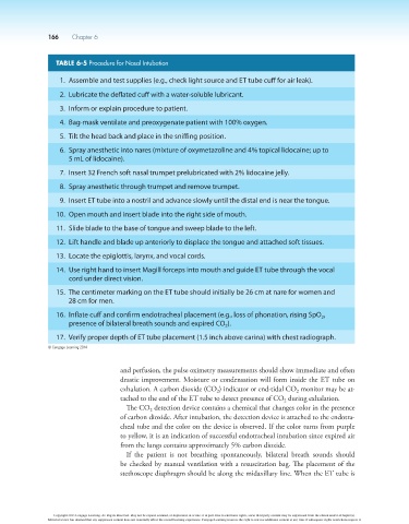
166    Chapter 6


                        TABLE 6-5 Procedure for Nasal Intubation

                          1.  Assemble and test supplies (e.g., check light source and ET tube cuff for air leak).

                          2.  Lubricate the deflated cuff with a water-soluble lubricant.

                          3.  Inform or explain procedure to patient.
                          4.  Bag-mask ventilate and preoxygenate patient with 100% oxygen.

                          5.  Tilt the head back and place in the sniffing position.
                          6.   Spray anesthetic into nares (mixture of oxymetazoline and 4% topical lidocaine; up to
                            5 mL of lidocaine).

                          7.  Insert 32 French soft nasal trumpet prelubricated with 2% lidocaine jelly.
                          8.  Spray anesthetic through trumpet and remove trumpet.

                          9.  Insert ET tube into a nostril and advance slowly until the distal end is near the tongue.
                        10.  Open mouth and insert blade into the right side of mouth.

                        11.  Slide blade to the base of tongue and sweep blade to the left.
                        12.  Lift handle and blade up anteriorly to displace the tongue and attached soft tissues.

                        13.  Locate the epiglottis, larynx, and vocal cords.

                        14.   Use right hand to insert Magill forceps into mouth and guide ET tube through the vocal
                            cord under direct vision.

                        15.   The centimeter marking on the ET tube should initially be 26 cm at nare for women and
                            28 cm for men.
                        16.   Inflate cuff and confirm endotracheal placement (e.g., loss of phonation, rising SpO ,
                                                                                                        2
                            presence of bilateral breath sounds and expired CO ).
                                                                           2
                        17.  Verify proper depth of ET tube placement (1.5 inch above carina) with chest radiograph.
                      © Cengage Learning 2014


                                            and perfusion, the pulse oximetry measurements should show immediate and often
                                            drastic improvement. Moisture or condensation will form inside the ET tube on
                                            exhalation. A carbon dioxide (CO ) indicator or end-tidal CO  monitor may be at-
                                                                        2
                                                                                                2
                                            tached to the end of the ET tube to detect presence of CO  during exhalation.
                                                                                             2
                                             The CO  detection device contains a chemical that changes color in the presence
                                                    2
                                            of carbon dioxide. After intubation, the detection device is attached to the endotra-
                                            cheal tube and the color on the device is observed. If the color turns from purple
                                            to yellow, it is an indication of successful endotracheal intubation since expired air
                                            from the lungs contains approximately 5% carbon dioxide.
                                             If  the  patient  is  not  breathing  spontaneously,  bilateral  breath  sounds  should
                                            be checked by manual ventilation with a resuscitation bag. The placement of the
                                            stethoscope diaphragm should be along the midaxillary line. When the ET tube is


                        Copyright 2013 Cengage Learning. All Rights Reserved. May not be copied, scanned, or duplicated, in whole or in part. Due to electronic rights, some third party content may be suppressed from the eBook and/or eChapter(s).
                      Editorial review has deemed that any suppressed content does not materially affect the overall learning experience. Cengage Learning reserves the right to remove additional content at any time if subsequent rights restrictions require it.