
Page 201 - Clinical Application of Mechanical Ventilation
P. 201
Airway Management in Mechanical Ventilation 167
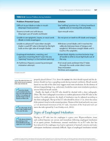
TABLE 6-6 Common Problems during Intubation
Problem (Potential Cause) Solution
Difficult to put blade or tube in mouth Use “sniffing” position by (1) tilting head back
(Improper head position) slightly and (2) moving chin anteriorly.
Trauma to teeth and soft tissues Open mouth wider.
(Improper use of handle and blade)
Unable to see epiglottis, larynx, or vocal cords Do not pivot on teeth to lift blade and tongue.
(Blade is inside esophagus)
Unable to advance ET tube when straight Withdraw curved blade until it reaches the
blade is used (ET tube is blocked by the light vallecula (between base of tongue and
bulb on the right side of straight blade) epiglottis). Withdraw straight blade until it
reaches the epiglottis.
Esophageal intubation, vomiting, and Rotate blade slightly counterclockwise (top
aspiration (Inserting the ET tube into any of handle to left) to move light bulb out of
“opening” hoping it is the tracheal opening) the way.
Arrhythmias (Hypoxia caused by prolonged Find vocal cords and insert the ET tube
intubation attempt) through the cords under direct vision.
Stop intubation.
Ventilate and oxygenate.
© Cengage Learning 2014
properly placed (about 1.5 in. above the carina) the chest should expand and the ab-
carina: The point at the lower end
of the trachea separating openings domen should not have a gurgling sound during manual ventilation. Breath sounds
of the main-stem bronchi. heard on one side of the chest may suggest main-stem intubation. In the absence of
obvious lung pathology (e.g., atelectasis), borderline main-stem intubation produces
uneven bilateral breath sounds.
Do not check breath
sounds at anterior chest loca- Finally, the depth of the ET tube should be checked with a chest radiograph.
tions close to the trachea since
airflow in the esophagus can (Note: The chest radiograph is not done to confirm placement of the ET tube in the
give false “breath sounds” in trachea.) For adult patients, the depth of intubation may be adjusted according to
neonates and thin adults.
the chest radiograph. The tip of the ET tube should be about 1.5 in. above the carina
if the patient’s head is in the neutral position. Flexion of the head and neck can cause
a 2 cm downward movement of the ET tube. Extension of the head and neck can
For adult patients, the tip move the tube upward by 2 cm (Godoy et al., 2012).
of an ET tube should be about
1.5 in. above the carina.
Signs of Esophageal Intubation
Placing an ET tube into the esophagus is a grave error. Hypoventilation, tissue
In the absence of obvious and cerebral hypoxia are certain and immediate following esophageal intubation
lung pathology, uneven
bilateral breath sounds may of an apneic patient. Furthermore, manual ventilation via an ET tube that has
suggest main-stem intubation. been placed in the esophagus may lead to aspiration of stomach contents and make
subsequent intubations extremely difficult. Signs of esophageal intubation include
Copyright 2013 Cengage Learning. All Rights Reserved. May not be copied, scanned, or duplicated, in whole or in part. Due to electronic rights, some third party content may be suppressed from the eBook and/or eChapter(s).
Editorial review has deemed that any suppressed content does not materially affect the overall learning experience. Cengage Learning reserves the right to remove additional content at any time if subsequent rights restrictions require it.