
Page 296 - Clinical Application of Mechanical Ventilation
P. 296
262 Chapter 9
A
Sensor Unit
Cell
ET Tube
Ventilator
B
Used with permission from Criticare Systems, Inc.
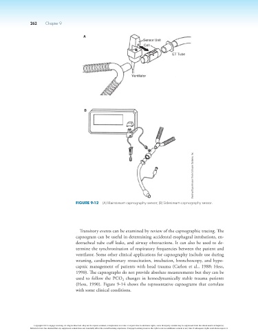
Figure 9-12 (A) Mainstream capnography sensor; (B) Sidestream capnography sensor.
Transitory events can be examined by review of the capnographic tracing. The
capnogram can be useful in determining accidental esophageal intubations, en-
dotracheal tube cuff leaks, and airway obstructions. It can also be used to de-
termine the synchronization of respiratory frequencies between the patient and
ventilator. Some other clinical applications for capnography include use during
weaning, cardiopulmonary resuscitation, intubation, bronchoscopy, and hypo-
capnic management of patients with head trauma (Carlon et al., 1988; Hess,
1990). The capnographs do not provide absolute measurements but they can be
used to follow the PCO changes in hemodynamically stable trauma patients
2
(Hess, 1990). Figure 9-14 shows the representative capnograms that correlate
with some clinical conditions.
Copyright 2013 Cengage Learning. All Rights Reserved. May not be copied, scanned, or duplicated, in whole or in part. Due to electronic rights, some third party content may be suppressed from the eBook and/or eChapter(s).
Editorial review has deemed that any suppressed content does not materially affect the overall learning experience. Cengage Learning reserves the right to remove additional content at any time if subsequent rights restrictions require it.