
Page 319 - Clinical Application of Mechanical Ventilation
P. 319
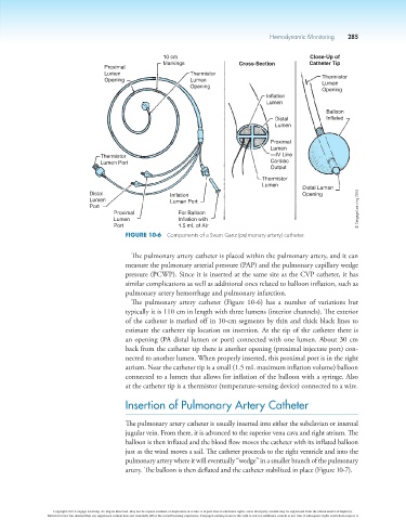
Hemodynamic Monitoring  285


                                                         10 cm                                       Close-Up of
                                                         Markings               Cross-Section        Catheter Tip
                                       Proximal
                                       Lumen                     Thermistor
                                       Opening                   Lumen                                   Thermistor
                                                                                                         Lumen
                                                                 Opening
                                                                                                         Opening
                                                                                        Inflation
                                                                                        Lumen
                                                                                                          Balloon
                                                                                           Distal         Inflated
                                                                                           Lumen

                                                                                         Proximal
                                                                                         Lumen
                                      Thermistor                                         —IV Line
                                      Lumen Port                                         Cardiac
                                                                                         Output
                                                                                       Thermistor
                                                                                       Lumen
                                                                                                   Distal Lumen
                                   Distal                  Inflation                               Opening
                                   Lumen                   Lumen Port
                                   Port                                                                             © Cengage Learning 2014
                                          Proximal           For Balloon
                                          Lumen              Inflation with
                                          Port               1.5 mL of Air
                                             Figure 10-6  Components of a Swan-Ganz (pulmonary artery) catheter.

                                               The pulmonary artery catheter is placed within the pulmonary artery, and it can
                                             measure the pulmonary arterial pressure (PAP) and the pulmonary capillary wedge
                                             pressure (PCWP). Since it is inserted at the same site as the CVP catheter, it has
                                             similar complications as well as additional ones related to balloon inflation, such as
                                             pulmonary artery hemorrhage and pulmonary infarction.
                                               The  pulmonary  artery  catheter  (Figure  10-6)  has  a  number  of  variations  but
                                             typically it is 110 cm in length with three lumens (interior channels). The exterior
                                             of the catheter is marked off in 10-cm segments by thin and thick black lines to
                                             estimate the catheter tip location on insertion. At the tip of the catheter there is
                                             an opening (PA distal lumen or port) connected with one lumen. About 30 cm
                                             back from the catheter tip there is another opening (proximal injectate port) con-
                                             nected to another lumen. When properly inserted, this proximal port is in the right
                                             atrium. Near the catheter tip is a small (1.5 mL maximum inflation volume) balloon
                                             connected to a lumen that allows for inflation of the balloon with a syringe. Also
                                             at the catheter tip is a thermistor (temperature-sensing device) connected to a wire.

                                             Insertion of Pulmonary Artery Catheter


                                             The pulmonary artery catheter is usually inserted into either the subclavian or internal
                                             jugular vein. From there, it is advanced to the superior vena cava and right atrium. The
                                             balloon is then inflated and the blood flow moves the catheter with its inflated balloon
                                             just as the wind moves a sail. The catheter proceeds to the right ventricle and into the
                                             pulmonary artery where it will eventually “wedge” in a smaller branch of the pulmonary
                                             artery. The balloon is then deflated and the catheter stabilized in place (Figure 10-7).


                        Copyright 2013 Cengage Learning. All Rights Reserved. May not be copied, scanned, or duplicated, in whole or in part. Due to electronic rights, some third party content may be suppressed from the eBook and/or eChapter(s).
                      Editorial review has deemed that any suppressed content does not materially affect the overall learning experience. Cengage Learning reserves the right to remove additional content at any time if subsequent rights restrictions require it.