
Page 433 - Clinical Application of Mechanical Ventilation
P. 433
Management of Mechanical Ventilation 399
Ventilator-Associated Pneumonia
Patients who are intubated and on mechanical ventilation are more prone to de-
velop nosocomial pneumonia than nonintubated patients (Craven et al., 1989). The
estimated incidence of ventilator-associated pneumonia (VAP) ranges from 10%
ventilator-associated
pneumonia (VAP): Infection to 65%, with fatality rates of 13% to 55% (Kollef et al., 1994). The presence of an
of the lung parenchyma that is artificial airway bypasses the natural defense mechanism of the airway, causes local
related to any or multiple events
that the patient undergoes during trauma and inflammation, and increases the risk of aspiration of pathogens from
mechan ical ventilation. the oropharynx.
In one study, 45% of the patients developed pneumonia within 3 days of intuba-
tion (Lowy et al., 1987). This condition may be caused by microbes acquired from
the patient’s oropharynx, respiratory instruments, health care providers (Hu, 1991),
endotracheal and nasogastric tubes (Joshi et al., 1993), and manual ventilation bags
(Weber et al., 1990). Table 12-11 outlines the potential sources of ventilator-associated
pneumonia. Strategies to decrease ventilator-associated pneumonias include proper
handwashing techniques, closed suction systems (Figure 12-6), continuous-feed
humidification systems, change of ventilator circuit only when visibly soiled, and
elevation of head of bed to 30° to 45° (Tablan et al., 2004).
For the diagnosis and treatment of VAP, early microbiologic examinations are
recommended to guide the use of appropriate antibiotics. Diagnosis and treatment
recommendations are beyond the scope of this chapter. Readers should research
current publications on VAP and read the articles by Rello et al. (2001) and Koenig
et al. (2006). Chapter 15 provides a more detailed discussion on VAP.
Sputum Culture. Sputum cultures should be obtained if infection of the lungs
is suspected. Since the patient is intubated, the sputum sample may be ob-
tained via an endotracheal suction setup and a sputum trap (Figure 12-7).
Sputum analyses are commonly done by the Gram stain, and the culture and
Gram stain: A method for
staining bacteria. Gram- positive sensitivity methods.
bacteria (e.g., Staphlococcus)
retain the gentian violet (purple)
color and gram-negative bacteria
(e.g., Pseudomonas) take the red
counterstain.
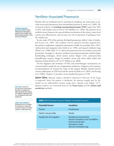
TABLE 12-11 Potential Sources of Ventilator-Associated Pneumonia
culture and sensitivity: A
laboratory procedure that grows Potential Source Locations
the microbes in a medium and
tests their sensitivity or resistance
to different antimicrobial drugs. Patient Oropharynx
Health care provider Hands
Equipment and supplies Respiratory instruments
Aerosol nebulizers and humidifiers
Endotracheal tube
Nasogastric tube
Manual ventilation bag
© Cengage Learning 2014
Copyright 2013 Cengage Learning. All Rights Reserved. May not be copied, scanned, or duplicated, in whole or in part. Due to electronic rights, some third party content may be suppressed from the eBook and/or eChapter(s).
Editorial review has deemed that any suppressed content does not materially affect the overall learning experience. Cengage Learning reserves the right to remove additional content at any time if subsequent rights restrictions require it.