
Page 466 - Clinical Application of Mechanical Ventilation
P. 466
432 Chapter 13
Since serious adverse effects can occur with the use of these agents, they should
not be given routinely or before alternative management is considered. If paralysis is
indicated, a sedative drug, such as the benzodiazepines along with an opioid analgesic,
should be provided for patient comfort. This is necessary because perception and pain
thresholds of a patient still exist with use of neuromuscular blocking drugs.
The following testimonies affirm the need for adequate sedation and analgesia
during paralysis. One patient who was pharmacologically paralyzed but not sedated
described his experience as “a feeling of being buried alive.” Another patient thought
that she had died (Halloran, 1991). A trauma survivor recalls the sensation of en-
dotracheal tube suctioning being like that of a red-hot burning iron passed into the
trachea (Hansen-Flaschen et al., 1993).
Mechanism of Action
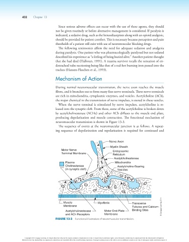
During normal neuromuscular transmission, the nerve axon reaches the muscle
fibers, and it branches out to form many fine nerve terminals. These nerve terminals
are rich in mitochondria, cytoplasmic enzymes, and vesicles. Acetylcholine (ACh),
the major chemical in the transmission of nerve impulses, is stored in these vesicles.
When the nerve terminal is stimulated by nerve impulses, acetylcholine is re-
leased into the synaptic cleft. From there, some of the acetylcholine is broken down
by acetylcholinesterase (ACHe) and other ACh diffuses to the muscle end plate,
producing depolarization and muscle contraction. The functional mechanism of
neuromuscular transmission is shown in Figure 13-3.
The sequence of events at the neuromuscular junction is as follows. A repeat-
ing sequence of depolarization and repolarization is required for continued and
Nerve Axon
Myelin Sheath
Motor Nerve Endoplasmic
Terminal Membrane Reticulum
Acetylcholinesterase
Plasma Mitochondria
Cholinesterase Acetylcholine-Bearing
(in synaptic cleft) Vesicles
Muscle Myofibrils Transverse
Membrane Tubules and Calcium- © Cengage Learning 2014
Acetylcholinesterase Motor End-Plate Binding Sites
and ACh Receptors Membrane
Figure 13-3 Functional illustration of neuromuscular transmission.
Copyright 2013 Cengage Learning. All Rights Reserved. May not be copied, scanned, or duplicated, in whole or in part. Due to electronic rights, some third party content may be suppressed from the eBook and/or eChapter(s).
Editorial review has deemed that any suppressed content does not materially affect the overall learning experience. Cengage Learning reserves the right to remove additional content at any time if subsequent rights restrictions require it.