
Page 48 - Clinical Application of Mechanical Ventilation
P. 48
14 Chapter 1
lower lung zone (more perfusion) to 3.0 in the upper lung zone (less perfusion)
(West, 2008).
In disease conditions, pulmonary embolism decreases pulmonary perfusion and
leads to a high V/Q. Airway obstruction is one example that leads to decrease in
ventilation and low V/Q.
V/Q mismatch is responsible for the development of hypoxemia. With sufficient
pulmonary reserve, a patient can usually compensate for the hypoxemic condition
by hyperventilation. Hypoxemia caused by uncomplicated V/Q mismatch is readily
reversible by oxygen therapy.
In mechanical ventilation, hypoxemia caused by V/Q mismatch can be compen-
sated by increasing the frequency, tidal volume, or F O on the ventilator (Shapiro
2
I
et al., 1991).
Intrapulmonary Shunting
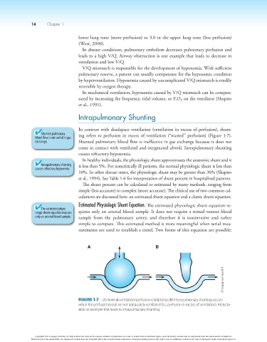
In contrast with deadspace ventilation (ventilation in excess of perfusion), shunt-
Shunted pulmonary ing refers to perfusion in excess of ventilation (“wasted” perfusion) (Figure 1-7).
blood flow is not useful in gas
exchange. Shunted pulmonary blood flow is ineffective in gas exchange because it does not
come in contact with ventilated and oxygenated alveoli. Intrapulmonary shunting
causes refractory hypoxemia.
In healthy individuals, the physiologic shunt approximates the anatomic shunt and it
Intrapulmonary shunting is less than 5%. For noncritically ill patients, the normal physiologic shunt is less than
causes refractory hypoxemia.
10%. In other disease states, the physiologic shunt may be greater than 30% (Shapiro
et al., 1994). See Table 1-6 for interpretation of shunt percent in hospitalized patients.
The shunt percent can be calculated or estimated by many methods, ranging from
simple (less accurate) to complex (more accurate). The clinical use of two common cal-
culations are discussed here: an estimated shunt equation and a classic shunt equation.
Estimated Physiologic Shunt Equation. The estimated physiologic shunt equation re-
The estimated physi-
ologic shunt equation requires quires only an arterial blood sample. It does not require a mixed venous blood
only an arterial blood sample. sample from the pulmonary artery, and therefore it is noninvasive and rather
simple to compute. This estimated method is more meaningful when serial mea-
surements are used to establish a trend. Two forms of this equation are possible:
A B
© Cengage Learning 2014
Figure 1-7 (A) Normal ventilation/perfusion relationship; (B) Intra-pulmonary shunting occurs
when the perfused alveoli are not adequately ventilated (i.e., perfusion in excess of ventilation). Atelecta-
sis is an example that leads to intrapulmonary shunting.
Copyright 2013 Cengage Learning. All Rights Reserved. May not be copied, scanned, or duplicated, in whole or in part. Due to electronic rights, some third party content may be suppressed from the eBook and/or eChapter(s).
Editorial review has deemed that any suppressed content does not materially affect the overall learning experience. Cengage Learning reserves the right to remove additional content at any time if subsequent rights restrictions require it.