
Page 46 - Clinical Application of Mechanical Ventilation
P. 46
12 Chapter 1
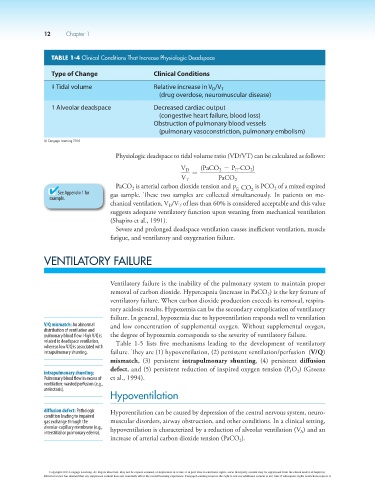
TABLE 1-4 Clinical Conditions That Increase Physiologic Deadspace
Type of Change Clinical Conditions
Relative increase in V /V T
➞ Tidal volume
D
(drug overdose, neuromuscular disease)
Alveolar deadspace Decreased cardiac output
➞
(congestive heart failure, blood loss)
Obstruction of pulmonary blood vessels
(pulmonary vasoconstriction, pulmonary embolism)
© Cengage Learning 2014
Physiologic deadspace to tidal volume ratio (VD/VT) can be calculated as follows:
V D = (PaCO - P -CO )
2
2
E
V T PaCO 2
PaCO is arterial carbon dioxide tension and P -CO is PCO of a mixed expired
2
2
E
2
See Appendix 1 for gas sample. These two samples are collected simultaneously. In patients on me-
example.
chanical ventilation, V /V of less than 60% is considered acceptable and this value
T
D
suggests adequate ventilatory function upon weaning from mechanical ventilation
(Shapiro et al., 1991).
Severe and prolonged deadspace ventilation causes inefficient ventilation, muscle
fatigue, and ventilatory and oxygenation failure.
VENTILATORY FAILURE
Ventilatory failure is the inability of the pulmonary system to maintain proper
removal of carbon dioxide. Hypercapnia (increase in PaCO ) is the key feature of
2
ventilatory failure. When carbon dioxide production exceeds its removal, respira-
tory acidosis results. Hypoxemia can be the secondary complication of ventilatory
failure. In general, hypoxemia due to hypoventilation responds well to ventilation
V/Q mismatch: An abnormal and low concentration of supplemental oxygen. Without supplemental oxygen,
distribution of ventilation and
pulmonary blood flow. High V/Q is the degree of hypoxemia corresponds to the severity of ventilatory failure.
related to deadspace ventilation, Table 1-5 lists five mechanisms leading to the development of ventilatory
whereas low V/Q is associated with
intrapulmonary shunting. failure. They are (1) hypoventilation, (2) persistent ventilation/perfusion (V/Q)
mismatch, (3) persistent intrapulmonary shunting, (4) persistent diffusion
defect, and (5) persistent reduction of inspired oxygen tension (P O ) (Greene
intrapulmonary shunting: I 2
Pulmonary blood flow in excess of et al., 1994).
ventilation; wasted perfusion (e.g.,
atelectasis).
Hypoventilation
diffusion defect: Pathologic Hypoventilation can be caused by depression of the central nervous system, neuro-
condition leading to impaired
gas exchange through the muscular disorders, airway obstruction, and other conditions. In a clinical setting,
alveolar-capillary membrane (e.g., hypoventilation is characterized by a reduction of alveolar ventilation (V ) and an
interstitial or pulmonary edema). A
increase of arterial carbon dioxide tension (PaCO ).
2
Copyright 2013 Cengage Learning. All Rights Reserved. May not be copied, scanned, or duplicated, in whole or in part. Due to electronic rights, some third party content may be suppressed from the eBook and/or eChapter(s).
Editorial review has deemed that any suppressed content does not materially affect the overall learning experience. Cengage Learning reserves the right to remove additional content at any time if subsequent rights restrictions require it.