
Page 45 - Clinical Application of Mechanical Ventilation
P. 45
Principles of Mechanical Ventilation 11
Anatomic Deadspace
Normally, the conducting airways contribute to about 30% of deadspace ventila-
anatomic deadspace: The
volume occupying the conducting tion. For a tidal volume of 500 mL, about 150 mL of this volume is wasted since it
airways that does not take part in does not take part in gas exchange. This volume in the conducting airways is called
gas exchange (estimated to be
1 mL/lb ideal body weight). anatomic deadspace and it can be estimated to be 1 mL/lb of ideal body weight
(Shapiro et al., 1991).
Decrease in tidal volume causes a relatively higher anatomic deadspace to tidal
Decrease in tidal volume volume percent. For example, if the tidal volume was decreased from 500 to 300 mL,
causes a relatively higher
anatomic deadspace to tidal the deadspace to tidal volume percent would increase from 30% (150/500) to 50%
volume percent. (150/300) See equations below for comparison:
150
500 = 0.3 or 30%
150 = 0.5 or 50%
300
Alveolar Deadspace
In addition to anatomic deadspace, alveolar deadspace may occur in some clinical con-
alveolar deadspace: The
normal lung volume that has ditions. Alveolar deadspace contributes to wasted ventilation, and it occurs when the
become unable to take part in gas ventilated alveoli are not adequately perfused by pulmonary circulation. Pulmonary per-
exchange because of reduction or
lack of pulmonary perfusion (e.g., fusion may be absent or low because of decreased cardiac output (e.g., congestive heart
pulmonary embolism).
failure, blood loss), or due to obstruction of the pulmonary blood vessels (e.g., pulmonary
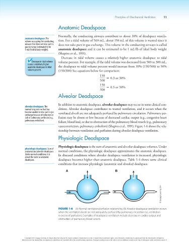
vasoconstriction, pulmonary embolism) (Shapiro et al., 1991). Figure 1-6 shows the rela-
tionship between ventilation and perfusion during alveolar deadspace ventilation.
Physiologic Deadspace
Physiologic deadspace is the sum of anatomic and alveolar deadspace volumes. Under
physiologic deadspace: Sum of
anatomic and alveolar deadspace. normal conditions, the physiologic deadspace approximates the anatomic deadspace.
Under normal conditions, it is In diseased conditions where alveolar deadspace ventilation is increased, physiologic
about the same as anatomic
deadspace. deadspace becomes higher than anatomic deadspace. Table 1-4 shows some clinical
conditions that increase physiologic (anatomic and alveolar) deadspace.
A B
© Cengage Learning 2014
Figure 1-6 (A) Normal ventilation/perfusion relationship; (B) Alveolar deadspace ventilation occurs
when the ventilated alveoli are not adequately perfused by pulmonary circulation (i.e., ventilation
in excess of perfusion). Examples of deadspace ventilation include decrease in cardiac output and
obstruction of pulmonary blood vessels.
Copyright 2013 Cengage Learning. All Rights Reserved. May not be copied, scanned, or duplicated, in whole or in part. Due to electronic rights, some third party content may be suppressed from the eBook and/or eChapter(s).
Editorial review has deemed that any suppressed content does not materially affect the overall learning experience. Cengage Learning reserves the right to remove additional content at any time if subsequent rights restrictions require it.