
Page 51 - Clinical Application of Mechanical Ventilation
P. 51
Principles of Mechanical Ventilation 17
Hypoxemia and Hypoxia
Hypoxemia is present when the oxygen level (e.g., PO , SaO ) is decreased in arte-
2
2
rial blood. The presence of hypoxia (TPO in organs and tissues) may not be always
2
apparent. Hypoxemia reflects the likelihood of hypoxia, but hypoxia can occur
in the absence of hypoxemia. For example, anemic hypoxia caused by reduced or
dysfunctional hemoglobins (e.g., anemia, blood loss, carbon monoxide poisoning),
histotoxic hypoxia caused by tissue dysfunction (e.g., cyanide poisoning), and cir-
culatory hypoxia caused by perfusion defects (e.g., Tcardiac output) may show nor-
mal PaO measurements (Shapiro et al., 1991).
2
Hypoxemia. Hypoxemia is reduced oxygen in the blood. The PaO from arterial
2
blood gases is commonly used to evaluate a patient’s oxygenation status. Since
PaO is a measurement of dissolved oxygen in the plasma, it does not represent
2
the portion of oxygen carried by the hemoglobin. For precise assessment, arte-
rial oxygen content (CaO ) measured by co-oximetry should be used because it
2
includes the oxygen combined with hemoglobin and the oxygen dissolved in the
plasma.
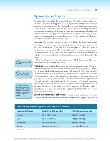
When PaO is used for oxygenation assessment, Table 1-8 may be used for inter-
2
Hypoxia can occur with a pretation of an adult’s oxygenation status.
normal PaO 2 .
Hypoxia. Hypoxia is reduced oxygen in the body organs and tissues. While hy-
poxemia and hypoxia are two terms sometimes used interchangeably, it is impor-
tant to understand that hypoxia can occur with a normal PaO . Two examples
2
CaO 2 5 (Hb 3 1.34 3 illustrate this point. Cyanide poisoning causes histotoxic hypoxia in which the
SaO 2 ) 1 (PaO 2 3 0.003)
tissues cannot carry out aerobic metabolism. Anemia causes a low oxygen content
(CaO ) while the PaO is often normal. Since PaO measures the oxygen tension
2
2
2
of the plasma only, it cannot be used for the assessment of histotoxic or anemic
hypoxia. CaO should be measured and used to assess the oxygenation status of
2
Oxygenation failure a patient. In addition to the PaO and CaO measurements, hypoxia produces
may develop when severe 2 2
hypoxemia (PaO 2 , 40 mm clinical signs (e.g., cyanosis, tachycardia, diaphoresis) that may be used as a sec-
Hg) does not respond to a ondary assessment tool.
moderate to high level (50%
to 100%) of supplemental
oxygen. Signs of Oxygenation Failure and Hypoxia. In most clinical situations, hypoxemia
is readily corrected by a moderate amount of supplemental oxygen. Oxygenation
TABLE 1-8 Interpretation of Oxygenation Status Using PaO 2 at Different P B
Hypoxemia Status PaO (P 5 760 mm Hg) PaO (P 5 630 mm Hg)
2
B
2
B
Normal 80 to 100 mm Hg 60 to 79 mm Hg
Mild 60 to 79 mm Hg 50 to 59 mm Hg
Moderate 40 to 59 mm Hg 40 to 49 mm Hg
Severe Less than 40 mm Hg Less than 40 mm Hg
© Cengage Learning 2014
Copyright 2013 Cengage Learning. All Rights Reserved. May not be copied, scanned, or duplicated, in whole or in part. Due to electronic rights, some third party content may be suppressed from the eBook and/or eChapter(s).
Editorial review has deemed that any suppressed content does not materially affect the overall learning experience. Cengage Learning reserves the right to remove additional content at any time if subsequent rights restrictions require it.