
Page 504 - Clinical Application of Mechanical Ventilation
P. 504
470 Chapter 14
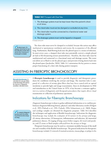
TABLE 14-1 Transport with Chest Tube
1. The drainage system must be kept lower than the patient’s chest
at all times.
2. The chest tube must not be clamped or occluded at any time.
3. The chest tube must be connected to a functional water seal
drainage system.
4. The drainage system must not be tipped or dropped.
© Cengage Learning 2014
The chest tube must never be clamped or occluded, because this action may affect
During transport, the mechanical or spontaneous ventilation and reverse the re-expansion of the affected
drainage system must be kept
lower than the patient’s chest lung. Furthermore, fluid flowing back into the pleural space is a source of infection.
and the chest tube must not In most severe cases, a clamped chest tube may potentially convert a simple pleural
be clamped or occluded.
air leak to life-threatening tension pneumothorax. Therefore, the chest tube must be
unobstructed and connected to a functional water seal drainage system. The water
seal allows air or fluid to exit the pleural space and prevents it being drawn back into
the pleural space (Jacobsohn, 2004). Table 14-1 summarizes the key points to ensure
proper functioning of a chest tube during patient transport.
ASSISTING IN FIBEROPTIC BRONCHOSCOPY
A fiberoptic bronchoscope is used to provide diagnostic and therapeutic proce-
fiberoptic bronchoscope: An
instrument that uses glass fibers dures for conditions involving the airways and lungs. The insertion tube is com-
to transmit images of the airway posed of a collection of minute glass fibers that have been coated with an optical
for diagnostic or therapeutic
procedures under direct vision. insulation to provide light and image transmission. Since its development in 1966
and introduction to the United States in 1970, it has become a common applica-
tion to a variety of diagnostic and therapeutic procedures that require direct visual
examination or collection of pulmonary specimens.
Indications for Fiberoptic Bronchoscopy
Diagnostic bronchoscopy is done to gather additional information or to confirm pre-
liminary diagnosis following history, physical, and other laboratory results (Holgate
et al., 1992; Prakash et al., 1991; Prakash, 1994; Schuurmans et al., 2003). Biopsy
and cytology samples are obtained for diagnostic purposes. Table 14-2 describes
An example of diagnostic the techniques and application of flexible bronchoscopy. Indications for diagnostic
bronchoscopy is the evalua-
tion of tumors in the airways bronchoscopy may include the evaluation of (1) tumors in the airways and lungs,
and lungs.
(2) airway obstruction, (3) hemoptysis, inflammation and infection, (4) interstitial
pulmonary disease, (5) staging of lung cancer before surgery, (6) vocal cord paraly-
sis, and (7) tissue or fluid samples collected from the airways or lungs.
Therapeutic bronchoscopy may be used as a treatment modality because of the small
size and versatility of the flexible bronchoscope. The general indications for therapeutic
bronchoscopy include (1) removal of retained secretions, mucus plugs, or polyps in the
Copyright 2013 Cengage Learning. All Rights Reserved. May not be copied, scanned, or duplicated, in whole or in part. Due to electronic rights, some third party content may be suppressed from the eBook and/or eChapter(s).
Editorial review has deemed that any suppressed content does not materially affect the overall learning experience. Cengage Learning reserves the right to remove additional content at any time if subsequent rights restrictions require it.