
Page 193 - Cardiac Nursing
P. 193
LWBK340-c07_p153-176.qxd 6/29/09 10:14 PM Page 169 Aptara Inc.
C HAP TE R 7 / Fluid and Electrolyte and Acid–Base Balance and Imbalance 169
peripheral vascular resistance and the decreased cardiac contractility metabolic acidosis by each of these mechanisms. Cardiogenic
can cause hypotension, which may be diminished by constriction in shock causes metabolic acidosis by accumulation of lactic acid
splanchnic and peripheral venous beds (the venous capacitance from anaerobic metabolism and through failure of the decreased
beds). This response increases central arterial blood volume. circulation to deliver metabolic acids to the kidneys for excretion.
The decreased pH in the CSF increases the synthesis of nitric No matter what its cause, metabolic acidosis is characterized by a
oxide, which causes cerebral vasodilation, increasing cerebral decreased plasma bicarbonate ion concentration. The bicarbonate
blood flow. 146 This is the source of the headache that is experi- either is depleted by being used to buffer excess metabolic acids or
enced by many individuals with respiratory acidosis. Increased is lost directly from the body.
cerebral blood flow from cerebral vasodilation may also raise CSF Some clinicians use the anion gap when evaluating metabolic
pressure and cause papilledema. In contrast to its effect on other acidosis. 148 The anion gap is the difference between the concen-
vascular beds, respiratory acidosis causes vasoconstriction in the trations of the major positive and negative ions in plasma or serum:
pulmonary vasculature. 144,147 The resulting increase in pul- Anion gap (Na
K ) (Cl
HCO 3 )
monary vascular resistance may worsen the clinical status of peo-
ple with preexisting right heart failure. Some people omit the potassium concentration, a relatively small
In summary, the major cardiovascular effects of respiratory aci- number, from the calculation to simplify it. The normal range of
dosis are tachycardia, cardiac arrhythmias, decreased cardiac con- anion gap varies with the laboratory procedures used for elec-
tractility, decreased peripheral vascular resistance, increased trolyte measurements, so that it may be reported as 6 to
pulmonary vascular resistance, and shift of blood flow from the ve- 16 mEq/L, 12 to 20 mEq/L, or another such range. 148–150 If un-
nous capacitance beds into the central and cerebral arterial beds. measured anions such as lactate or -hydroxybutyrate accumulate
in the body, the anion gap increases. Calculation of the anion gap
Metabolic Acidosis is rapid and uses clinically available parameters, but it is less in-
Metabolic acidosis is caused by relatively too much metabolic formative for individuals who have hypoalbuminemia unless a
acid. It can be due to a gain of acid or a loss of base. 139 Acid can correction is used and may be misleading when two primary
be gained from intake of acids or substances that are converted to acid–base imbalances coexist. 150
acid in the body, from an increased rate of normal metabolism, Calculating the anion gap enables division of metabolic acido-
from production of unusual acids due to altered metabolic sis into two groups: high serum anion gap metabolic acidosis and
processes, or from factors that decrease renal excretion of acid. Bi- normal serum anion gap metabolic acidosis. 3,148 The anion gap in-
carbonate ions (base) can be lost in the urine or through the gas- creases when an abnormal metabolic acid accumulates in the
trointestinal tract. Table 7-18 lists clinical conditions that cause body, such as with lactic acidosis or ketoacidosis. Normal anion
gap acidosis, also called hyperchloremic acidosis, typically occurs
with diarrhea or loss of HCO 3 from the kidneys, which retain
NaCl in response. Critically ill patients who have lactic acidosis, a
type of high anion gap acidosis, have been shown to have a higher
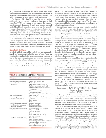
Table 7-18 ■ CAUSES OF METABOLIC ACIDOSIS mortality rate than those who have normal anion gap acidosis. 151
In research and some clinical settings, metabolic acid–base im-
Category Clinical Examples
balances may be evaluated using a quantitative physical chemistry
Acid accumulation by ingestion Aspirin (acetylsalicylic acid) method often called the Stewart approach or the strong ion gap
or infusion of acid or acid Boric acid (SIG). 151,152 The SIG is the apparent strong ion difference minus
precursors Ammonium chloride (releases H )
Methanol (converts to formic acid) the charge on buffer base:
Antifreeze (ethylene glycol converts to Strong ion gap (Na
K
Caa a 2
Mgg g 2
Cl lactate)
oxalic acid)
Paraldehyde (converts to acetic and (charge on albumin
charge on
chloroacetic acids) phosphate
HCO 3 )
Elemental sulfur (converts to sulfuric acid)
Acid accumulation by Hyperthyroidism One advantage of the SIG over the traditional anion gap is that it
increased production of Hypermetabolic state after burns, trauma, enables quantification of the unmeasured anion, but disadvantages
normal metabolic acids or sepsis are that the calculation is time-consuming and includes clinical pa-
Lactic acidosis rameters such as serum magnesium and lactate concentrations that
Shock 153
y
Acid accumulation by utilization Alcoholic ketoacidosis frequently are not readily available. Whether the SIG is useful in
151–153
of abnormal or incomplete Diabetic ketoacidosis predicting outcomes of metabolic acidosis is controversial.
metabolic pathways Starvation ketoacidosis Metabolic acidosis can be corrected physiologically only by the
Acid accumulation by impaired Prolonged oliguria from any cause kidneys, which are the sole excretory route for metabolic acids.
acid exceration Oliguric renal failure Renal correction of metabolic acidosis may take several days.
Severe hypovolemia
Shock Meanwhile, respiratory compensation occurs within hours. The
Renal tubular acidosis (type 1) respiratory compensation for metabolic acidosis is hyperventilation.
Hypoaldosteronism By increasing the excretion of carbonic acid, hyperventilation
Loss of base (bicarbonate ions) Severe diarrhea makes the blood less acid. This makes the blood chemistry more ab-
Intestinal decompression
Fistula drainage from pancreas or intestine normal (decreased Pa CO2 ), but tends to restore the 20:1 ratio of
Vomiting of intestinal contents bicarbonate to carbonic acid and move the pH toward the normal
Ureterosigmoidostomy range, thus helping to preserve cellular function. Compensated
Renal tubular acidosis (type 2) metabolic acidosis is characterized by a decreased Pa CO2 (the sign
of the respiratory compensation), a decreased bicarbonate ion