
Page 194 - Cardiac Nursing
P. 194
LWBK340-c07_p153-176.qxd 6/29/09 10:14 PM Page 170 Aptara Inc.
170 PA R T II / Physiologic and Pathologic Responses
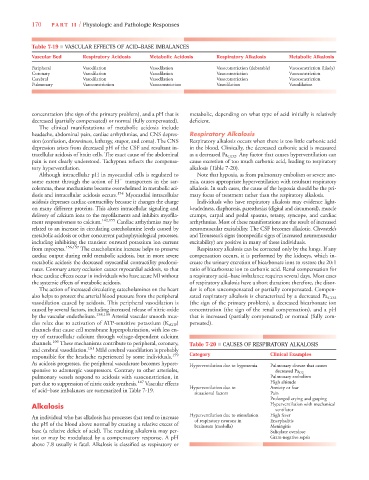
Table 7-19 ■ VASCULAR EFFECTS OF ACID–BASE IMBALANCES
Vascular Bed Respiratory Acidosis Metabolic Acidosis Respiratory Alkalosis Metabolic Alkalosis
Peripheral Vasodilation Vasodilation Vasoconstriction (debatable) Vasoconstriction (likely)
Coronary Vasodilation Vasodilation Vasoconstriction Vasoconstriction
Cerebral Vasodilation Vasodilation Vasoconstriction Vasoconstriction
Pulmonary Vasoconstriction Vasoconstriction Vasodilation Vasodilation
concentration (the sign of the primary problem), and a pH that is metabolic, depending on what type of acid initially is relatively
decreased (partially compensated) or normal (fully compensated). deficient.
The clinical manifestations of metabolic acidosis include
headache, abdominal pain, cardiac arrhythmias, and CNS depres- Respiratory Alkalosis
sion (confusion, drowsiness, lethargy, stupor, and coma). The CNS Respiratory alkalosis occurs when there is too little carbonic acid
depression arises from decreased pH of the CSF and resultant in- in the blood. Clinically, the decreased carbonic acid is measured
tracellular acidosis of brain cells. The exact cause of the abdominal as a decreased Pa CO2 . Any factor that causes hyperventilation can
pain is not clearly understood. Tachypnea reflects the compensa- cause excretion of too much carbonic acid, leading to respiratory
tory hyperventilation. alkalosis (Table 7-20).
Although intracellular pH in myocardial cells is regulated to Note that hypoxia, as from pulmonary embolism or severe ane-
some extent through the action of H
transporters in the sar- mia, causes appropriate hyperventilation with resultant respiratory
colemma, these mechanisms become overwhelmed in metabolic aci- alkalosis. In such cases, the cause of the hypoxia should be the pri-
dosis and intracellular acidosis occurs. 154 Myocardial intracellular mary focus of treatment rather than the respiratory alkalosis.
acidosis depresses cardiac contractility because it changes the charge Individuals who have respiratory alkalosis may evidence light-
on many different proteins. This alters intracellular signaling and headedness, diaphoresis, paresthesias (digital and circumoral), muscle
delivery of calcium ions to the myofilaments and inhibits myofila- cramps, carpal and pedal spasms, tetany, syncope, and cardiac
ment responsiveness to calcium. 142,155 Cardiac arrhythmias may be arrhythmias. Most of these manifestations are the result of increased
related to an increase in circulating catecholamine levels caused by neuromuscular excitability. The CSF becomes alkalotic. Chvostek’s
metabolic acidosis or other concurrent pathophysiological processes, and Trousseau’s signs (nonspecific signs of increased neuromuscular
including inhibiting the transient outward potassium ion current excitability) are positive in many of these individuals.
from myocytes. 156,157 The catecholamine increase helps to preserve Respiratory alkalosis can be corrected only by the lungs. If any
cardiac output during mild metabolic acidosis, but in more severe compensation occurs, it is performed by the kidneys, which in-
metabolic acidosis the decreased myocardial contractility predomi- crease the urinary excretion of bicarbonate ions to restore the 20:1
nates. Coronary artery occlusion causes myocardial acidosis, so that ratio of bicarbonate ion to carbonic acid. Renal compensation for
these cardiac effects occur in individuals who have acute MI without a respiratory acid–base imbalance requires several days. Most cases
the systemic effects of metabolic acidosis. of respiratory alkalosis have a short duration; therefore, the disor-
The action of increased circulating catecholamines on the heart der is often uncompensated or partially compensated. Compen-
also helps to protect the arterial blood pressure from the peripheral sated respiratory alkalosis is characterized by a decreased Pa CO2
vasodilation caused by acidosis. This peripheral vasodilation is (the sign of the primary problem), a decreased bicarbonate ion
caused by several factors, including increased release of nitric oxide concentration (the sign of the renal compensation), and a pH
by the vascular endothelium. 158,159 Arterial vascular smooth mus- that is increased (partially compensated) or normal (fully com-
cles relax due to activation of ATP-sensitive potassium (K ATP ) pensated).
K
channels that cause cell membrane hyperpolarization, with less en-
try of extracellular calcium through voltage-dependent calcium
channels. 160 These mechanisms contribute to peripheral, coronary, Table 7-20 ■ CAUSES OF RESPIRATORY ALKALOSIS
and cerebral vasodilation. 161 Mild cerebral vasodilation is probably
responsible for the headache experienced by some individuals. 159 Category Clinical Examples
As acidosis progresses, the peripheral vasculature becomes hypore- Hyperventilation due to hypoxemia Pulmonary disease that causes
sponsive to adrenergic vasopressors. Contrary to other arterioles, decreased Pa O2
pulmonary vessels respond to acidosis with vasoconstriction, in Pulmonary embolism
part due to suppression of nitric oxide synthesis. 147 Vascular effects High altitude
of acid–base imbalances are summarized in Table 7-19. Hyperventilation due to Anxiety or fear
situational factors Pain
Prolonged crying and gasping
Alkalosis Hyperventilation with mechanical
ventilator
An individual who has alkalosis has processes that tend to increase Hyperventilation due to stimulation High fever
of respiratory neurons in
Encephalitis
the pH of the blood above normal by creating a relative excess of brainstem (medulla) Meningitis
base (a relative deficit of acid). The resulting alkalemia may per- Salicylate overdose
sist or may be modulated by a compensatory response. A pH Gram-negative sepsis
above 7.8 usually is fatal. Alkalosis is classified as respiratory or