Page 243 - Cardiac Nursing
P. 243

0:4
                                        6 A
                                     1
                                      0:4
                                          M
                                            Pa
                                        6 A
                                          M
                                     1
                              0/0
                              0/0
                             3
                             3
                                  009
                                  009
                                6/2
                                6/2
                                                   p
                                                   p
                                                  A
                                                   p
                                                     ara
                                                     ara
                                                    t
                                                    t
                                                  A
                                              g
                                              g
                                            Pa
                                              g
                                                19
                                                19
                                               e 2
                                               e 2
                           xd
                      1-2
         LWBK340-c10_
            K34
         LWB K34 0-c 10_ pp211-244.qxd  30/06/2009  10:46 AM  Page 219 Aptara
         LWB
                      1-2
                    21
                    21
                   p
               0-c
                 10_
                   p
                          q
                        44.
                          q
                           xd
                          q
                        44.
                                                               C HAPTER 1 0 / History Taking and Physical Examination  219
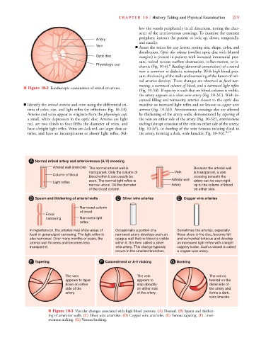
                                                                        low the vessels peripherally in all directions, noting the char-
                                                                        acter of the arteriovenous crossings. To examine the extreme
                                                                        periphery, instruct the patient to look up, down, temporally,
                                                    Artery
                                                                        and nasally.
                                                    Vein               ■ Assess the retina for any lesions, noting size, shape, color, and
                                                                        distribution. Optic disc edema (swollen optic disc with blurred
                                                    Optic disc          margins) is present in patients with increased intracranial pres-
                                                                        sure, retinal venous outflow obstruction, inflammation, or is-
                                                    Physiologic cup                   8
                                                                        chemia (Fig. 10-4). Beading (abnormal constriction) of a retinal
                                                                        vein is common in diabetic retinopathy. With high blood pres-
                                                                        sure, thickening of the walls and narrowing of the lumen of reti-
                                                                        nal arteries develop. These changes are observed as focal nar-
                                                                        rowing, a narrowed column of blood, and a narrowed light reflex
                   ■ Figure 10-2 Funduscopic examination of retinal structures.
                                                                        (Fig. 10-3B). If opacity is such that no blood column is visible,
                                                                                B
                                                                        the artery appears as a silver wire artery (Fig. 10-3C). With in-
                                                                        creased filling and tortuosity, arteries closest to the optic disc
                   ■ Identify the retinal arteries and veins using the differential cri-  manifest an increased light reflex and are known as copper wire
                     teria of color, size, and light reflex (or reflection; Fig. 10-3A).  arteries (Fig. 10-3D). Arteriovenous crossings also are affected
                     Arteries and veins appear to originate from the physiologic cup,  by thickening of the artery walls, demonstrated by tapering of
                     a small, white depression in the optic disc. Arteries are light  the vein on either side of the artery (Fig. 10-3E), arteriovenous
                     red, are two thirds to four fifths the diameter of veins, and  nicking (abrupt cessation of the vein on either side of the artery;
                     have a bright light reflex. Veins are dark red, are larger than ar-  Fig. 10-3F), or banking of the vein (venous twisting distal to
                     teries, and have an inconspicuous or absent light reflex. Fol-  the artery, forming a dark, wide knuckle; Fig. 10-3G). 8,17
                      A  Normal retinal artery and arteriovenous (A-V) crossing
                                Arterial wall (invisible)  The normal arterial wall is            Because the arterial wall
                                                 transparent. Only the column of         Vein     is transparent, a vein
                                Column of blood
                                                 blood within it can usually be                   crossing beneath the
                                                 seen. The normal light reflex is      Arterial wall  artery can be seen right
                                Light reflex
                                                 narrow–about 1/4 the diameter         Artery     up to the column of blood
                                                 of the blood column.                             on either side.
                      B  Spasm and thickening of arterial walls  C  Silver wire arteries  D  Copper wire arteries
                                             Narrowed column
                                             of blood
                             Focal
                             narrowing       Narrowed light
                                             reflex
                      In hypertension, the arteries may show areas of   Occasionally a portion of a   Sometimes the arteries, especially
                      focal or generalized narrowing. The light reflex is   narrowed artery develops such an   those close to the disc, become full
                      also narrowed. Over many months or years, the   opaque wall that no blood is visible   and somewhat tortuous and develop
                      arterial wall thickens and becomes less   within it. It is then called a silver   an increased light reflex with a bright
                                                                    y
                                                                    y
                      transparent.                           wire artery. This change typically   coppery luster. Such a vessel is called
                                                                                                       y
                                                                                                       y
                                                             occurs in the smallest branches.   a copper wire artery.
                      E  Tapering                     F  Concealment or A-V nicking    G  Banking
                                      The vein                         The vein                          The vein is
                                      appears to taper                 appears to                        twisted on the
                                      down on either                   stop abruptly                     distal side of
                                      side of the                      on either side                    the artery and
                                      artery.                          of the artery.                    forms a dark,
                                                                                                         wide knuckle.
                              ■ Figure 10-3 Vascular changes associated with high blood pressure. (A) Normal. (B) Spasm and thicken-
                              ing of arteriolar walls. (C) Silver wire arterioles. (D) Copper wire arterioles. (E) Venous tapering. (F) Arteri-
                              ovenous nicking. (G) Venous banking.
   238   239   240   241   242   243   244   245   246   247   248