
Page 247 - Cardiac Nursing
P. 247
6/2
009
6/2
0/0
0/0
0:4
0:4
1
009
1
3
44.
q
44.
1-2
1-2
xd
3
xd
q
q
6 A
p
p
A
23
A
ara
ara
t
p
t
23
Pa
Pa
M
6 A
M
e 2
e 2
g
g
g
p
LWB K34 0-c 10_ p pp211-244.qxd 30/06/2009 10:46 AM Page 223 Aptara
21
21
LWB
0-c
10_
LWBK340-c10_
K34
C HAPTER 1 0 / History Taking and Physical Examination 223
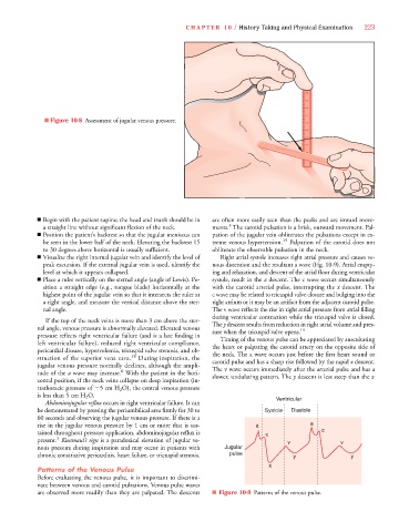
■ Figure 10-8 Assessment of jugular venous pressure.
■ Begin with the patient supine; the head and trunk should be in are often more easily seen than the peaks and are inward move-
3
a straight line without significant flexion of the neck. ments. The carotid pulsation is a brisk, outward movement. Pal-
■ Position the patient’s backrest so that the jugular meniscus can pation of the jugular vein obliterates the pulsations except in ex-
be seen in the lower half of the neck. Elevating the backrest 15 treme venous hypertension. 19 Palpation of the carotid does not
to 30 degrees above horizontal is usually sufficient. obliterate the observable pulsation in the neck.
■ Visualize the right internal jugular vein and identify the level of Right atrial systole increases right atrial pressure and causes ve-
peak excursion. If the external jugular vein is used, identify the nous distention and the resultant a wave (Fig. 10-9). Atrial empty-
level at which it appears collapsed. ing and relaxation, and descent of the atrial floor during ventricular
■ Place a ruler vertically on the sternal angle (angle of Lewis). Po- systole, result in the x descent. The c wave occurs simultaneously
sition a straight edge (e.g., tongue blade) horizontally at the with the carotid arterial pulse, interrupting the x descent. The
highest point of the jugular vein so that it intersects the ruler at c wave may be related to tricuspid valve closure and bulging into the
a right angle, and measure the vertical distance above the ster- right atrium or it may be an artifact from the adjacent carotid pulse.
nal angle. The v wave reflects the rise in right atrial pressure from atrial filling
during ventricular contraction while the tricuspid valve is closed.
If the top of the neck veins is more than 3 cm above the ster- The y descent results from reduction in right atrial volume and pres-
nal angle, venous pressure is abnormally elevated. Elevated venous sure when the tricuspid valve opens. 19
pressure reflects right ventricular failure (and is a late finding in Timing of the venous pulse can be appreciated by auscultating
left ventricular failure), reduced right ventricular compliance, the heart or palpating the carotid artery on the opposite side of
pericardial disease, hypervolemia, tricuspid valve stenosis, and ob- the neck. The a wave occurs just before the first heart sound or
struction of the superior vena cava. 18 During inspiration, the carotid pulse and has a sharp rise followed by the rapid x descent.
jugular venous pressure normally declines, although the ampli- The v wave occurs immediately after the arterial pulse and has a
6
tude of the a wave may increase. With the patient in the hori- slower, undulating pattern. The y descent is less steep than the x
zontal position, if the neck veins collapse on deep inspiration (in-
trathoracic pressure of 5 cm H 2 O), the central venous pressure
is less than 5 cm H 2 O. Ventricular
Abdominojugular reflux occurs in right ventricular failure. It can
be demonstrated by pressing the periumbilical area firmly for 30 to Systole Diastole
60 seconds and observing the jugular venous pressure. If there is a
rise in the jugular venous pressure by 1 cm or more that is sus- a a
tained throughout pressure application, abdominojugular reflux is c c
3
present. Kussmaul’s sign is a paradoxical elevation of jugular ve- v v
nous pressure during inspiration and may occur in patients with Jugular
chronic constrictive pericarditis, heart failure, or tricuspid stenosis. pulse y y
x
Patterns of the Venous Pulse
Before evaluating the venous pulse, it is important to discrimi-
nate between venous and carotid pulsations. Venous pulse waves
are observed more readily than they are palpated. The descents ■ Figure 10-9 Patterns of the venous pulse.