
Page 244 - Cardiac Nursing
P. 244
xd
xd
q
q
q
3
6/2
6/2
0/0
3
0/0
p
21
t
ara
t
21
44.
44.
1-2
p
1-2
g
e 2
g
Pa
g
e 2
A
p
A
20
20
1
0:4
1
009
009
0:4
M
Pa
M
6 A
6 A
ara
K34
0-c
LWBK340-c10_
LWB
10_
LWB K34 0-c 10_ p p pp211-244.qxd 30/06/2009 10:46 AM Page 220 Aptara
220 P A R T III / Assessment of Heart Disease
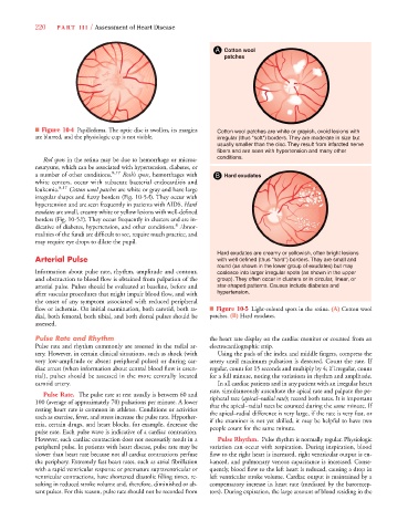
A Cotton wool
patches
■ Figure 10-4 Papilledema. The optic disc is swollen, its margins Cotton wool patches are white or grayish, ovoid lesions with
are blurred, and the physiologic cup is not visible. irregular (thus "soft") borders. They are moderate in size but
usually smaller than the disc. They result from infarcted nerve
fibers and are seen with hypertension and many other
Red spots in the retina may be due to hemorrhage or microa- conditions.
neurysms, which can be associated with hypertension, diabetes, or
a number of other conditions. 8,17 Roth’s spots, hemorrhages with B Hard exudates
white centers, occur with subacute bacterial endocarditis and
leukemia. 8,17 Cotton wool patches are white or gray and have large
irregular shapes and fuzzy borders (Fig. 10-5A). They occur with
hypertension and are seen frequently in patients with AIDS. Hard
exudates are small, creamy white or yellow lesions with well-defined
borders (Fig. 10-5B). They occur frequently in clusters and are in-B
B
8
dicative of diabetes, hypertension, and other conditions. Abnor-
malities of the fundi are difficult to see, require much practice, and
may require eye drops to dilate the pupil.
Hard exudates are creamy or yellowish, often bright lesions
Arterial Pulse with well defined (thus "hard") borders. Thay are small and
round (as shown in the lower group of exudates) but may
Information about pulse rate, rhythm, amplitude and contour, coalesce into larger irregular spots (as shown in the upper
and obstruction to blood flow is obtained from palpation of the group). They often occur in clusters or in circular, linear, or
arterial pulse. Pulses should be evaluated at baseline, before and star-shaped patterns. Causes include diabetes and
after vascular procedures that might impair blood flow, and with hypertension.
the onset of any symptom associated with reduced peripheral
flow or ischemia. On initial examination, both carotid, both ra- ■ Figure 10-5 Light-colored spots in the retina. (A) Cotton wool
dial, both femoral, both tibial, and both dorsal pulses should be patches. (B) Hard exudates.
assessed.
Pulse Rate and Rhythm the heart rate display on the cardiac monitor or counted from an
Pulse rate and rhythm commonly are assessed in the radial ar- electrocardiographic strip.
tery. However, in certain clinical situations, such as shock (with Using the pads of the index and middle fingers, compress the
very low-amplitude or absent peripheral pulses) or during car- artery until maximum pulsation is detected. Count the rate. If
diac arrest (when information about central blood flow is essen- regular, count for 15 seconds and multiply by 4; if irregular, count
tial), pulses should be assessed in the more centrally located for a full minute, noting the variations in rhythm and amplitude.
carotid artery. In all cardiac patients and in any patient with an irregular heart
rate, simultaneously auscultate the apical rate and palpate the pe-
Pulse Rate. The pulse rate at rest usually is between 60 and ripheral rate (apical–radial rate); record both rates. It is importante e
100 (average of approximately 70) pulsations per minute. A lower that the apical–radial rates be counted during the same minute. If
resting heart rate is common in athletes. Conditions or activities the apical–radial difference is very large, if the rate is very fast, or
such as exercise, fever, and stress increase the pulse rate. Hypother- if the examiner is not yet skilled, it may be helpful to have two
mia, certain drugs, and heart blocks, for example, decrease the people count for the same minute.
pulse rate. Each pulse wave is indicative of a cardiac contraction.
However, each cardiac contraction does not necessarily result in a Pulse Rhythm. Pulse rhythm is normally regular. Physiologic
peripheral pulse. In patients with heart disease, pulse rate may be variation can occur with respiration. During inspiration, blood
slower than heart rate because not all cardiac contractions perfuse flow to the right heart is increased, right ventricular output is en-
the periphery. Extremely fast heart rates, such as atrial fibrillation hanced, and pulmonary venous capacitance is increased. Conse-
with a rapid ventricular response or premature supraventricular or quently, blood flow to the left heart is reduced, causing a drop in
ventricular contractions, have shortened diastolic filling times, re- left ventricular stroke volume. Cardiac output is maintained by a
sulting in reduced stroke volume and, therefore, diminished or ab- compensatory increase in heart rate (mediated by the barorecep-
sent pulses. For this reason, pulse rate should not be recorded from tors). During expiration, the large amount of blood residing in the