
Page 245 - Cardiac Nursing
P. 245
3
3
xd
q
xd
0/0
009
009
6/2
0/0
6/2
21
ara
21
t
ara
1-2
q
q
44.
1-2
44.
1
21
21
e 2
g
e 2
A
p
t
p
A
p
6 A
6 A
0:4
1
0:4
M
g
g
Pa
M
Pa
0-c
10_
10_
0-c
LWBK340-c10_ p p pp211-244.qxd 30/06/2009 10:46 AM Page 221 Aptara
K34
K34
C HAPTER 1 0 / History Taking and Physical Examination 221
pulmonary vascular bed during inspiration reaches the left heart.
Left ventricular contractility is enhanced by means of the Frank– mm Hg
Starling mechanism, increasing left ventricular stroke volume. Be- A
cause an increased heart rate is no longer needed to maintain car-
diac output, the heart rate returns to baseline. This physiologically
irregular rhythm is termed sinus arrhythmia. It is common in peo- B
ple younger than 40 years of age. Other irregular rhythms are not
normal. The irregularity should be described as regularly irregular
(e.g., every other pulse wave is early) or irregularly irregular (e.g.,
atrial fibrillation). Occasional, early pulsations that are perceived as
transient skips or breaks in an otherwise regular rhythm are com- C
mon and are not necessarily abnormal.
Pulse Amplitude and Contour D
Pulses are described in a variety of ways. The simplest classifica-
tion is absent, present, and bounding. A 0-to-4 scale is often used,
and pulses are graded as follows: absent (0), diminished (1
),
normal (2
), moderately increased (3
), and markedly increased
18
(4
). This scale is fairly subjective, and, although an individual E
tends to be internally consistent over time, different people may
grade the same pulse differently. There are also other scales in
which the numbers are defined differently.
The amplitude of an arterial pulse is a function of the pulse F
pressure, which is related to stroke volume, elasticity of the arte- Premature contractions
rial tree, and velocity of left ventricular ejection. Increased stroke
volume, as occurs with exercise or excitement, results in increased
amplitude and a bounding arterial pulse. G
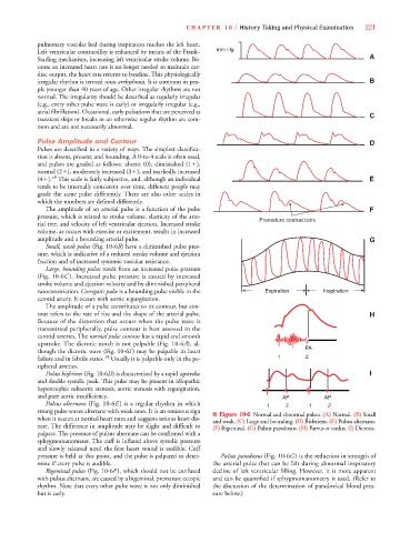
Small, weak pulses (Fig. 10-6B) have a diminished pulse pres-B
sure, which is indicative of a reduced stroke volume and ejection
fraction and of increased systemic vascular resistance.
Large, bounding pulses result from an increased pulse pressure
(Fig. 10-6C ). Increased pulse pressure is caused by increased
stroke volume and ejection velocity and by diminished peripheral
vasoconstriction. Corrigan’s pulse is a bounding pulse visible in the Expiration Inspiration
carotid artery. It occurs with aortic regurgitation.
The amplitude of a pulse contributes to its contour, but con-
tour refers to the rate of rise and the shape of the arterial pulse. H
Because of the distortion that occurs when the pulse wave is
transmitted peripherally, pulse contour is best assessed in the
carotid arteries. The normal pulse contour has a rapid and smooth
upstroke. The dicrotic notch is not palpable (Fig. 10-6A), al-
6
6
though the dicrotic wave (Fig. 10-6I ) may be palpable in heart PA
18
failure and in febrile states. Usually it is palpable only in the pe- 1 2
ripheral arteries.
Pulsus bisferiens (Fig. 10-6D) is characterized by a rapid upstroke I
and double systolic peak. This pulse may be present in idiopathic
hypertrophic subaortic stenosis, aortic stenosis with regurgitation,
and pure aortic insufficiency. AP AP
Pulsus alternans (Fig. 10-6E ) is a regular rhythm in which 1 2 1 2
strong pulse waves alternate with weak ones. It is an ominous sign ■ Figure 10-6 Normal and abnormal pulses. (A) Normal. (B) Small
when it occurs at normal heart rates and suggests serious heart dis- and weak. (C) Large and bounding. (D) Bisferiens. (E) Pulsus alternans.
ease. The difference in amplitude may be slight and difficult to (F) Bigeminal. (G) Pulsus paradoxus. (H) Parvus et tardus. (I) Dicrotic.
palpate. The presence of pulsus alternans can be confirmed with a
sphygmomanometer. The cuff is inflated above systolic pressure
and slowly released until the first heart sound is audible. Cuff
pressure is held at this point, and the pulse is palpated to deter- Pulsus paradoxus (Fig. 10-6G) is the reduction in strength of
mine if every pulse is audible. the arterial pulse that can be felt during abnormal inspiratory
Bigeminal pulses (Fig. 10-6F), which should not be confused decline of left ventricular filling. However, it is more apparent
with pulsus alternans, are caused by a bigeminal, premature ectopic and can be quantified if sphygmomanometry is used. (Refer to
rhythm. Note that every other pulse wave is not only diminished the discussion of the determination of paradoxical blood pres-
but is early. sure below.)