
Page 259 - Cardiac Nursing
P. 259
LWBK340-c10_p211-244.qxd 30/06/2009 10:47 AM Page 235 Aptara
C HAP TE R 1 0 / History Taking and Physical Examination 235
S 4 S 1 S 3 S 4 S 1 S 3 S 4 S 1 S 3
S
S
S
2
2
2
S 1 S OS S 1
2
■ Figure 10-21 Quadruple rhythm.
■ Figure 10-23 Opening snap (OS).
although one possibility may be an absence of actual mechanical
atrial contraction in spite of electrical atrial activity.
Opening snaps are associated with the opening of a stenotic mi-
tral valve. Opening sounds are not heard with normal valves. The
sound is heard in very early diastole, medial to the cardiac apex. S 1 E j S 2
The sound can be loud and transmitted throughout the pre-
cordium (Fig. 10-23). Unlike an S 3 , an opening snap has a high- ■ Figure 10-24 Early systolic ejection sound.
pitched, snapping quality and is heard best with the diaphragm of
the stethoscope. 33
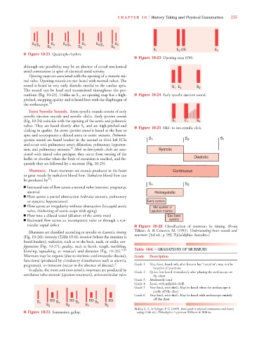
Extra Systolic Sounds. Extra systolic sounds consist of early
systolic ejection sounds and systolic clicks. Early ejection sounds
(Fig. 10-24) coincide with the opening of the aortic and pulmonic S 1 C 1 S 2
valves. They are heard shortly after S 1 and are high-pitched and ■ Figure 10-25 Mid- to late-systolic click.
clicking in quality. An aortic ejection sound is heard at the base or
apex and accompanies a dilated aorta or aortic stenosis. Pulmonic
ejection sounds are heard loudest in the second or third left ICSs S 1 S 2 S 1
and occur with pulmonary artery dilatation, pulmonary hyperten-
sion, and pulmonary stenosis. 33 Mid- to late-systolic clicks are asso- Systolic
ciated with mitral valve prolapse; they occur from tensing of the
leaflet or chordae when the limit of excursion is reached, and fre- Diastolic
quently they are followed by a murmur (Fig. 10-25).
Murmurs. Heart murmurs are sounds produced in the heart Continuous
or great vessels by turbulent blood flow. Turbulent blood flow can
33
be produced by :
S 1 S 2
■ Increased rate of flow across a normal valve (exercise, pregnancy,
anemia) Holosystolic
■ Flow across a partial obstruction (valvular stenosis, pulmonary
or systemic hypertension) Early systolic
■ Flow across an irregularity without obstruction (bicuspid aortic
Mid systolic or
valve, thickening of aortic cusps with aging) ejection murmur
■ Flow into a dilated vessel (dilation of the aortic root) End (late)
■ Backward flow across an incompetent valve or through a ven- systolic
tricular septal defect ■ Figure 10-26 Classification of murmurs by timing. (From
Murmurs are classified according to systolic or diastolic timing Tilkian, A. & Conover, M. [1993]. Understanding heart sounds and
(Fig. 10-26); intensity (Table 10-6); location (where the murmur is murmurs [3rd ed., p. 99]. Philadelphia: Saunders.)
heard loudest); radiation, such as to the back, neck, or axilla; con-
figuration (Fig. 10-27); quality, such as harsh, rough, rumbling,
blowing, squeaking, or musical; and duration (Fig. 10-26). 6,33 Table 10-6 ■ GRADATIONS OF MURMURS
Murmurs may be organic (due to intrinsic cardiovascular disease), Grade Description
functional (produced by circulatory disturbances such as anemia,
pregnancy), or innocent (occur in the absence of disease). 3 Grade 1 Very faint, heard only after listener has “tuned in”; may not be
heard in all positions
In adults, the most common systolic murmurs are produced by
Grade 2 Quiet, but heard immediately after placing the stethoscope on
semilunar valve stenosis (ejection murmurs), atrioventricular valve the chest
Grade 3 Moderately loud
Grade 4 Loud, with palpable thrill
Grade 5 Very loud, with thrill. May be heard when the stethoscope is
partly off the chest
Grade 6 Very loud, with thrill. May be heard with stethoscope entirely
off the chest
S 1 SG S 1 SG S 1 SG S 1 SG
S 2 S 2 S 2 S 2
Bickley, L. S., & Szilagyi, P. G. (2009). Bates’ guide to physical examination and history
■ Figure 10-22 Summation gallop. taking (10th ed.). Philadelphia: Lippincott Williams & Wilkins.