
Page 299 - Cardiac Nursing
P. 299
g
g
Pa
g
75
75
e 2
e 2
Pa
1:1
8 P
1
1:1
M
Pa
8 P
M
a
a
a
a
c.
c.
In
In
ara
p
p
A
A
t
ara
p
t
1
7-2
7-2
26
26
q
q
76.
76.
LWBK340-c12_
K34
LWB K34 0-c 12_ pp267-276.qxd 6/29/09 11:18 PM Page 275 Aptara Inc.
LWB
p
p
0-c
12_
/29
/29
6
1
/09
/09
/09
xd
xd
q
6
C HAPTER 12 / Radiologic Examination of the Chest 275
Small
thoracostomy
tube
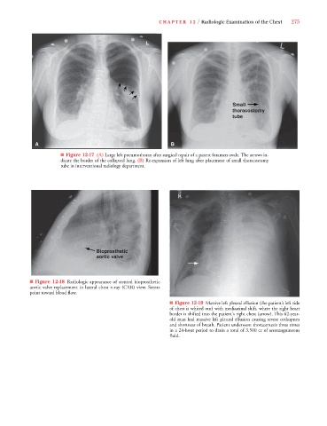
A B
■ Figure 12-17 (A) Large left pneumothorax after surgical repair of a patent foramen ovale. The arrows in-
dicate the border of the collapsed lung. (B) Re-expansion of left lung after placement of small thoracostomy
tube in interventional radiology department.
Bioprosthetic
aortic valve
■ Figure 12-18 Radiologic appearance of stented bioprosthetic
aortic valve replacement in lateral chest x-ray (CXR) view. Stents
point toward blood flow.
■ Figure 12-19 Massive left pleural effusion (the patient’s left side
of chest is whited out) with mediastinal shift, where the right heart
border is shifted into the patient's right chest (arrow). This 62-year-
old man had massive left pleural effusion causing severe orthopnea
and shortness of breath. Patient underwent thoracentesis three times
in a 24-hour period to drain a total of 3,500 cc of serosanguineous
fluid.