
Page 298 - Cardiac Nursing
P. 298
Pa
g
Pa
Pa
g
e 2
74
g
e 2
1
1:1
1
1
1:1
M
M
8 P
8 P
a
a
ara
a
a
c.
c.
In
In
A
p
74
A
p
t
ara
p
t
26
26
p
76.
7-2
7-2
p
LWBK340-c12_
LWB
LWB K34 0-c 12_ pp267-276.qxd 6/29/09 11:18 PM Page 274 Aptara Inc.
12_
0-c
K34
/09
/09
/09
6
/29
/29
6
q
q
76.
xd
xd
q
274 P A R T III / Assessment of Heart Disease
A
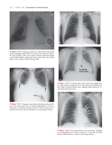
■ Figure 12-13 Cardiogenic pulmonary edema with cardiomegaly.
In this radiograph, Kerley B lines are seen as tiny horizontal lines in
the lung periphery. These lines represent dilated pulmonary lymph
vessels, which facilitate pulmonary edema removal from the alveolar
spaces. (Film courtesy of Julie Takasugi, MD)
Prosthetic
aortic valve
B
■ Figure 12-15 (A) Preoperative patient with aortic regurgitation.
(B) Same patient 3 weeks after aortic valve replacement. Patient now
has a large pericardial effusion (note enlarged cardiac silhouette) cre-
ating tamponade physiology.
■ Figure 12-14 Congestive heart failure with bilateral pleural effu-
sions. The arrows show that the normal diaphragmatic contour is
obliterated. This 88-year-old man with severe aortic stenosis was ad-
mitted with severe shortness of breath and worsening heart failure.
■ Figure 12-16 Descending thoracic aortic aneurysm. “Bulging”
of the descending aorta noted by arrows in a 57-year-old man with
history of blunt trauma to chest as well as hypertension.